11th Class

It is passage of metabolites, by-products and biochemicals across biomembrane. Membrane transport occurs through four methods–passive, facilitated, active and bulk. Size of the particles passing through plasmalemma is generally \[115\text{ }{AA}.\] Passive transport : No energy spent. Passive transport occurs through diffusion and osmosis. (1) Diffusion : It is movement of particles from the region of their higher concentration or electrochemical potential to the region of their lower concentration or electrochemical potential. Electrochemical potential operates in case of charged particles like ions. Simple diffusion does not require carrier molecules. (2) Osmosis : It is diffusion of water across a semipermeable membrane that occurs under the influence of an osmotically active solution. Mechanism of passive transport : Passive transport can continue to occur if the absorbed solute is immobilised. Cations have a tendency to passively pass from electropositive to electronegative side. While anions can pass from electronegative to electropositive side. There more...

Lysosomes are electron microscopic, vesicular structures of the cytoplasm, bounded by a single membrane (lipoproteinous) which are involved in intracellular digestive activities, contains hydrolytic enzymes, so called lysosomes. Discovery (i) These were first discovered by a Belgian biochemist, Christian de Duve (1955) in the liver cells and were earlier named pericanalicular dense bodies. (ii) Terms Lysosome was given by Novikoff under the study of electron microscope. (iii) Matile (1964) was first to demonstrate their presence in plants, particularly in the fungus Neurospora. Polymorphism in lysosomes were described by De Robertis et. al (1971). Occurrence : These are absent from the prokaryotes but are present in all eukaryotic animal cells except mammalian RBCs. They have been recorded in fungi, Euglena, cotton and pea seeds. Shape : These are generally spherical in shape but are irregular in plant root tip cells. Size : Size range is \[0.2-0.8\,\,\mu m\] while size is \[0.5\,\,\mu more...

Golgi complex is made up of various membranous system e.g., cisternae, vesicles and vacuoles. These are also called golgi bodies, golgisomes, lipochondrion, dictyosomes, Dalton complex, idiosomes or Baker’s body and “traffic police” of the cell. Discovery : First observed by George (1867) but it’s morphological details were given by Camillo Golgi (1898), in nerve cells of barn owl and cat. Occurence : It is present in all eukaryotic cells. In plants, these are scattered irregularly in the cytoplasm and called as “dictyosomes”. These are absent in bacteria and blue green algae, RBCs, spermatozoa of bryophytes and pteridophytes, and sieve tube cells of phloem of angiosperm. The number of golgi body increased during cell division. Average number 10 – 20 per cell. Golgi body surrounded by a zone of protoplasm which is devoid of cell organelles called zone of exclusion (Morre, 1977). Structure : Under transmission electron microscope the st. of more...

The substance occur around the nucleus and inside the plasma membrane containing various organelles and inclusions is called cytoplasm. (1) The cytoplasm is a semisolid, jelly – like material. It consists of an aqueous, structureless ground substance called cytoplasmic matrix or hyaloplasm or cytosol. (2) It forms about half of the cell’s volume and about 90% of it is water. (3) It contains ions, biomolecules, such as sugar, amino acid, nucleotide, tRNA, enzyme, vitamins, etc. (4) The cytosol also contains storage products such as glycogen/starch, fats and proteins in colloidal state. (5) It also forms crystallo – colloidal system. (6) Cytomatrix is differentiated into ectoplasm or plasmagel (outer) and endoplasm or plasmasol (inner). (7) Cytomatrix is three dimensional structure appear like a network of fine threads and these threads are called microfilaments (now called actin filaments or microtrabecular lattice) and it is believed to be a part of cytoskeleton. It more...

Discovery : Flagellum presence was first reported by Englemann (1868). Jansen (1887) was first scientist to report the structure of sperm flagellum. Definition : Cilia and flagella are microscopic, hair or thread-like motile structures present extra-cellularly but originate intra-cellularly from the basal body. Occurrence : Cilia are found in all the ciliate protozoans e.g., Paramecium, Vorticella etc. Flagella are found in all the flagellate protozoans e.g., Euglena, Trichonympha etc. Structure : Both cilia flagella are structurally similar and possess similar parts-basal body, rootlets, basal plate and shaft.     
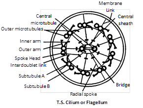
(1) Basal body : These are also termed as blepharoplast (kinetosome) or basal granule. It is present below the plasma membrane in cytoplasm. The structure is similar to centriole made of 9 triplets of microtubules. (2) Rootlets : Made of microfilament and providing support to the basal body. (3) Basal plate : Central more...

Discovery : Centrosome was first discovered by Van Benden (1887) and structure was given by T. Boweri. Occurrence : It is found in all the animal cell except mature mammalian RBC’s. It is also found in most of protists and motile plant cells like antherozoids of ferns, zoospores of algae and motile algal forms e.g., Chlamydomonas but is absent in prokaryotes, fungi, gymnosperms and angiosperms. Structure : Centrosome is without unit membrane structure. It is formed of two darkly stained granules called centrioles, which are collectively called diplosome. These centrioles are surrounded by a transparent cytoplasmic area called centrosphere of Kinetoplasm. Centriole and centrosphere are collectively called centrosome. Each centriole is a microtubular structure and is formed of microtubules arranged in 9+0 manner (all the 9 microtubules are peripheral in position). Inside the microtubules, there is an intra-centriolar or cart-wheel structure which is formed of a central hub (about \[25{AA}\] more...

Discovery : It was first discovered by Robert Hooke in 1665 in Cork. Cell wall is the outer most, rigid, protective, non living and supportive layer found in all the plant cells, bacteria, cyanobacteria and some protists. It is not found in animal cells. Chemical composition : Mainly cell wall consists of two parts, matrix and cellulosic fibres (microfibrils). Matrix consists of hemicellulose, pectin, glycoproteins, lipids and water.In most of the plants cell wall is made up of cellulose                                                                            \[{{({{C}_{6}}{{H}_{10}}{{O}_{5}})}_{n}},\] a polymer made-up of unbranched chain of glucose molecule linked by                                                                                                      \[\beta ,\,1-4\] glycosidic bond. About 100 molecules of cellulose form a micelle, about 20 micelle form a microfibril and approx 200 microfibril form a fibril. The cell wall of bacteria and the inner layer of blue green algae is made-up mucopeptide. It is a polymer of two amino sugars namely N-acetyl glucosamine (NAG) and N-acetyl muramic acid more...

Cytology : (Gk Kyios = cell ; logas = study) It is the branch of biology. Which comprises the study of cell structure and function. “Cell is the structural and functional unit of all living beings”. Study of metabolic aspects of cell components is called cell biology. Robert Hooke (1665) discovered hollow cavities (empty boxes) like compartments in a very thin slice of cork (cell wall) under his microscope. He wrote a book “Micrographia” and coined the term cellula, which was later changed into cell. Grew and Malpighi also observed small structures in slice of plants and animals. Leeuwenhoek was the first to see free cells and called them “wild animalcules” and published a book “The secret of nature”. He observed bacteria, protozoa, RBCs, sperms, etc. under his microscope. Cell theory : H.J. Dutrochet (1824) a French worker gave the idea of cell theory. The actual credit for cell theory more...

Mitochondria (Gk. Mito = thread ; chondrion = granule) are semi autonomous having hollow sac like structures present in all eukaryotes except mature RBCs of mammals and sieve tubes of phloem. Mesosomes of prokaryotes (bacteria) is analogous to mitochondrion in eukaryotes. Mitochondria are also called chondriosome, chondrioplast, plasmosomes, plastosomes and plastochondriane. Discoveries (1) These were first observed in striated muscles (Voluntary) of insects as granules by Kolliker (1880), he called them “sarcosomes”. (2) Flemming (1882) called them “fila” for thread like structure. (3) Altman (1890) called them “bioplast”. (4) C. Benda (1897) gave the term mitochondria. (5) F. Meves (1904) observed mitochondria in plant (Nymphaea). (6) Michaelis (1898) demonstrated that mitochondria play a significant role in respiration. (7) Bensley and Hoerr (1934) isolated mitochondria from liver cells. (8) Seekevitz called them “Power house of the cell”. (9) Nass and Afzelius (1965) observed first DNA in mitochondria. Number of mitochondria : more...

It is a technique of studying the route of chemicals in chemical reactions taking place inside the cell and organisms with the help of radioactive isotope. e.g., \[^{14}C,{{\,}^{3}}H,{{\,}^{32}}P.\] In this technique the radioisotopes are incorporated into the precursor molecule. Then the labelled precursor molecules introduced into the cells and their path is followed with the help of their radiations. Radioactive precursors emit radiations and their position in the cell is located by bringing the cell in contact with a photographic plate or film. \[^{32}P\]and \[^{14}C\] are used for the study of nucleic acids and photosynthesis (Melvin Calvin) respectively.


Archive


You need to login to perform this action.
You will be redirected in 3 sec spinner