11th Class

Origin and position : The thymus is bilobed gland, is located in the upper part of the thorax near the heart in the mediastinum. It is endodermal in origin, arising in the embryo from the epithelium of outer part of third branchial pouches. Structure : Structurally, it is like lymph gland enveloped by a thin, loose and fibrous connective tissue capsule. Septa, or trabeculae extending inwards from the capsule, divide the two lobes of the gland into a number of small lobules. Each lobule is distinguished into a cortical parenchyma containing numerous lymphocytes, and a medullary mass of large, irregularly branched and interconnected epithelial cells (reticular cells), a few lymphocytes and some phagocytic cells called macrophages or Hassal's corpuscles.     
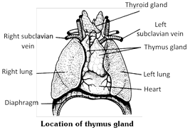
Function of thymus glands (1) Thymus is haemopoietic, as well as, an endocrine gland. Thymus more...

The name "thyroid" was introduced by Thomas Wharton (1656). It is derived from Greek "Thyreos" a shield. Location : This is the largest endocrine gland of our body. It is located in our neck upon the ventral aspect of larynx (sound box or Adam's apple) and a few anteriomost tracheal rings. It is a dark brown and H-shaped/butterfly bilobed gland. Origin : It is endodermal in origin and arises in the embryo as a midventral process from the floor of the tongue in pharyngeal region between the first and second pharyngeal pouches. Later, the duct-like connection (thyroglossal duct) of the process degenerates, so that the process is separated from the tongue and becomes endocrine. Probably, the gland is homologous to the endostyle of lower chordates. Structure of thyroid gland : In adult human beings, thyroid gland measures about 5 cm in length and 3 cm in width. It's average weight more...

Nervous system is divided into three parts -     
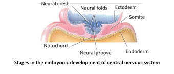
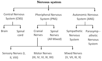
Central nervous system (CNS) In all the vertebrates including man, CNS is dorsal, hollow and non-ganglionated while in invertebrates when present, it is ventral, solid, double and ganglionated. CNS is formed of two parts : (1) Brain - Upper and broader part lying in the head. (2) Spinal cord - Lower, long and narrow part running from beginning of neck to trunk. CNS is covered by 3 meninges and its wall has two type of matter. Types of matter : CNS of vertebrates is formed of two types of matter – (i) Grey matter : It is formed of cell-bodies, non-medullated nerve fibres, neuroglea, dendrites of association neurons and motor neurons. (ii) White matter : It is formed of medullated nerve fibres or myelinated axon of more...

(1) Coelenterata : True nerve cell or ganglion cells occur for the first time in coelenterates. They are derived from interstitial cells of epidermis, forming nerve net or nerve plexus below whole epidermis. A polar neurons are found in coelenterata. (2) Platyhelminthes : Nervous system of planarians marks the beginning of a centralized nervous system encountered in higher animals. That is made up of brain or cerebral ganglia, two lateral longitudinal nerve chords, numerous peripheral nerves and transverse commissures or connectives. This is sometimes called the ladder type of nervous system. In Nematoda (e.g. ascaris) these system made up of central nervous system, peripheral nervous system and rectal nervous system. Rectal nervous system more developed in male. Ascaris with dorsal, ventral, and lateral nerve cords. (3) Annelida : Nervous system well developed and concentrated. It consists of three parts : central nervous system, peripheral nervous system and sympathetic nervous system, more...

Nervous system begins develop early in third week of development from ectoderm. Nervous tissue also develop from ectoderm except microglial cell, develop from mesoderm. The central nervous system of vertebrates includes the brain and the spinal cord. These are derived from a longitudinal mid-dorsal ectodermal thickening of the embryo, called the medullary or neural plate. This neural plate or neural groove is converted by fusion into a closed mid-dorsal longitudinal neural tube lying above the notochord. Histologically, the embryonic neural tube exhibits three zones of cells.         
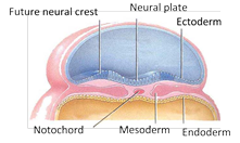
(1) Germinal layer : These are actively dividing cells lining the neural canal. They form the connective tissue more...

Nerve cells (= neurons) : Irritability is a basic characteristic of the “living substance”, i.e., the protoplasm. Consequently, every living cell becomes excited when stimulated. However, the nerve cells and muscle fibres are specialized excitable cells of body, capable of transmitting or conducting excitations along their membranes. Of these, muscle cells are further specialized for contraction while nerve cells are further specialized for receiving stimuli (as sensory or receptor cells) and transferring excitations from one to the other. A typical neuron consists of a nucleated cell body (= cyton, soma or perikaryon), five to seven short, slender and branched (= arborized) dendrites, and a single, relatively thicker and longer fibrous axon. The latter is terminally branched into short telodendria. Each telodendron bears a terminal knob or botton. Bottons of one neuron lie upon dendrites or cytons of adjacent neurons (figure), or upon muscle fibres or glands. Nerve fibres : Although, more...

The hardened tissues of the body together form the skeleton (sclero = hard). Skeleton of invertebrates is most often secreted on the surface, forming a lifeless or dead exoskeleton. Whereas skeleton of vertebrates develops most often underneath the surface forming a living or growing endoskeleton. Three types of skeletons develop in vertebrates : (1) Epidermal/Horny exoskeleton : These include hard and horny of keratinized derivatives of epidermal layer of skin, such as claws, most reptilian’s scales, bird feathers and mammalian hairs, horns, nails and hoofs, etc. All living amphibians lack an exoskeleton. (2) Dermal/Bony skeleton : Dermal bony skeleton is derived from the dermis of skin. It includes bony scales and plates or scutes (osteoderms), finrays and antlers of fishes, some reptiles (crocodiles, turtles and tortoises) and mammals. In fishes, dermal scales become exposed due to wearing out of epidermis, and form exoskeleton. (3) Endoskeleton : Greater part of vertebrate more...

Movement is one of the most important characteristics of living organisms. Nonliving objects do not move. If nonliving objects show movement, that is always due to some external force. For example, the cart is moved by the horse and the fan revolves by the energy of electric current. The movement of a nonliving object is, therefore induced (due to external force) while the movement of living things are autonomic (self sustained). Study of movement is called kinesiology (G. Kinein = to move, Logos = study). The movement of living systems are thus autonomic or active, that is effected by the organisms themselves without external influences. On the other hand the movement of nonliving systems are induced or passive, i.e., made to occur by external forces. Movement of animals are two main types muscular and non muscular. (1) Muscular movement : Muscular movement are found in the majority of animals brought more...

There are many articulations or joints present in the skeleton. Joint or articulations is a point of contact between bones. Joints are classified based upon their structure and the kinds of movements which they permit. Three main types of joints are - (1) Immovable joints (Synarthroses) : No joint cavity, no movement possible. These joints include - (i) Sutures : Found between skull bones, sutures are fixed or fibrous joints, articulating bones are held together by white fibrous tissue. (ii) Gomphoses : It is a type of fibrous joint in which cone shaped peg fits soket. Teeth in mandibles, and maxillary bones.     
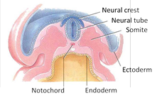
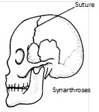
(iii) Syndesmosis : It is type of fibrous joint with more fibrous tissue than sutures. e.g., distal articulation between Tibia and fibula. (2) Imperfect joints (Amphiarthroses) slightly movable : Joints in which more...

(1) Axial endoskeleton : (Skull + Vertebral column + Sternum + Ribs) (2) Appendicular endoskeleton : (Girdle + Limb bones)       
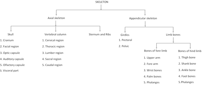
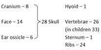
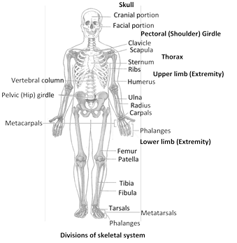
Axial skeleton (Human) It occupies the body's main longitudinal axis. It includes four structure : skull in the head, vertebral column in the neck, trunk and tail if present, sternum and ribs in the thorax. It form the upright axis of body and includes 80 (87 in children) bones are as follows in man -     (1) Skull (General structure) : It is anterior most axial skeleton. It is divisible into two main parts – (i) Chondrocranium                                         (ii) Splanchnocranium    (i) Chondrocranium : more...


Archive


You need to login to perform this action.
You will be redirected in 3 sec spinner