(1) Characteristics of female's skeleton : In female's skeleton, (i) skull is lighter, (ii) shoulders are narrower (iii) sacrum is shorter but wider, (iv) pelvis in wider, has a broader front and larger bottom opening to facilitate child birth, and (v) coccyx is more movable than in male's skeleton.
(2) Adaptations in skeleton for upright posture : Human skeleton shows many adaptive features for upright posture –
(i) Foramen magnum is directed downward so that the head may rest vertically on the vertebral column.
(ii) Four curves in the backbone keep the centre of gravity near the heels. This helps to maintain balance and makes walking erect on two legs much easier.
(iii) Thorax is wider from side-to-side than from front-to-back. This helps to maintain equilibrium.
(iv) Bowel-like pelvis supports the lower abdominal viscera.
(v) Metacarpals form a wide palm and the pollex is opposable. This make the hand a grasping organ to work with it.
(vi) Leg bones are stronger than the arm bones as the femur carry the entire weight of the body in locomotion.
(vii) Broad feet provide stability in the upright posture.
(viii) The arches of the feet enable the body to move with a degree of springiness.
(ix) Increased mobility of the neck to see all round.
(x) Increased skeletal height provides greater range of vision.
(3) Types of bones : Bones are divided into 4 categories regarding their size and shape –
(i) Long bones, e.g., humerus of upper arm, radius and ulna of forearm, femur of thigh, and tibia and fibula of leg.
(ii) Short bones, e.g., metacarpals of palm and metatarsals of foot, phalanges of fingers and toes.
(iii) Flat bones, e.g., scapula of shoulder girdle, sternum, cranial bones.
(iv) Irregular bones, e.g., vertebrae, carpals of wrist and tarsals of ankle.
(4) Bone movement : Movements of bones occur only at the joints. The movements are brought about by contractions of skeletal muscles inserted onto the articulating bones by firm cords of white fibrous tissue called tendons. Cords of yellow elastic tissue, termed ligaments, stabilise the joints by holding the articulating bones together.
(5) Disorder of skeleton and joints : Any violent movement, such as jump, fall or knock, may cause injury to the skeleton. The injury can be of 5 types - sprain, dislocation, fracture, arthritis and slipped disc.
(i) Sprain : Sprain refers to injury to a joint capsule, typically involving a stretching or tearing of tendons or ligaments. Unfortunately, both these structures have much poorer regenerative power than bone, and once stretched often remain weak. Sprain is often considered a minor disorder, but it may become chronic.
(ii) Arthritis or Aching Joints : Arthritis refers to inflammation of the joints. It is a common disease of the old age. Its common symptoms are pain and stiffness in the joints. It has many forms. Three more common forms are described here - osteoarthritis or degenerative arthritis, the rheumatoid arthritis and gout.
(a) Osteoarthritis : Secretion of the lubricating synovial fluid more...
In the body of all the multicellular animals muscles are found. The movement of the body takes place by these muscles. If the muscles become weak, the functioning of the body become difficult. The muscles are capable of contraction and relaxation, hence these are elastic.
A muscle can pull a part of the body by its contraction (shortening). It cannot push that part by relaxation (elongation). Hence, the muscles are typically arranged in antagonistic (opposing) pairs, one muscle moves a body part in one direction by its contraction and the other muscle moves that part in the opposite direction by its contraction. Of course, when one muscle contracts, its opposing muscle relaxes. The principle of antagonistic muscles is true of both vertebrate as well as invertebrate muscles. Animal movements depend upon interaction of muscles and skeleton.
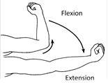
(1) Action of body muscles : As mentioned above, the body muscles are arranged in antagonistic (opposing) pairs. One muscle of a pair moves a body part in one direction and the other in the opposite direction. For example, the muscle named biceps brings the forearm toward the upper arm, and the muscle called the triceps moves the forearm away from the upper arm. When biceps contracts to cause movement, the triceps relaxes to allow that movement to occur and vice versa. Similar pairs of opposing flexor and extensor muscles occur at the wrist, ankle and knee. The type of movement that results from the contraction of a muscle depends entirely upon the way the muscle is attached to the levers of the skeleton.
(2) Classification of body muscles : According to the type of motion they cause, the muscles are divided into the following types. The muscles that act together to produce a movement are called synergists and the muscle that act in opposition to each other are antagonists. The muscles that act most powerfully during any given movements are called prime movers.
(i) Flexor and Extensor : Muscles that bend one part over another joint is called flexor. Extensor muscle is antagonist of flexor muscle. The contraction of an extensor extends a joint by pulling one of the articulating bone apart from another.
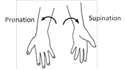
(ii) Pronator and Supinator : The contraction of a pronator rotates the forearm to turn the palm downward or backward. Supinator is antagonist of pronator. A supinator contracts to rotate the forearm and thus to make palm face upward or forward.
(iii) Abductor and Adductor : An abductor contracts to draw a bone away from the body midline. Muscle that brings the limb away from midline is called abductor. An adductor draws a bone towards the body midline. Muscles that brings the limb towards midline is called adductor. Abductor muscle is antagonist of adductor muscle. Abduction is elevation and adduction is depression.
more...
Circulatory system in various groups of animals can be classified as follows :
(1) Intracellular circulation : Occurs inside the individual cells where the distribution of substances is through cyclosis of cell cytoplasm. Example – Protozoans.
(2) Extracellular circulation : When the distribution of the substances occurs inside the body through extracellular or intracellular fluids. This is of following types –
(i) Extra organismic circulation : Canal system in porifera, water vascular system in Echinoderms and gastrovascular system in coelenterates.
(ii) Intra-organismic circulation : It involves circulation of body fluids. It is of following types –
(a) Parenchymal circulation : In platyhelminthes, the fluid filled spaces present in the mesodermal parenchyma tissue between body wall and internal organs are used in the distribution of substances.
(b) Coelomic circulation : Coelomic fluid is concerned with the transport of substances. Example – pseudocoelomic fluid in the roundworms and haemolymph in Arthropods.
(c) Blood vascular system : It contains blood and a pumping structure (heart) for circulation of materials inside the body. It is open circulatory system and closed circulatory system.
Differences between open and closed circulatory system
S.N.
Open circulatory system
Closed circulatory system
1.
In open circulatory system blood flows through large open spaces and channels called lacunae and sinuses among the tissues.
In closed circulatory system blood flows through a closed system of chambers called heart and blood vessels.
2.
Tissues are in direct contact with the blood.
Blood does not come in direct contact with tissue.
It is a part of venous circulation which is present between two groups of capillaries i.e. starts in capillaries and ends in capillaries. The vein which drains blood into organs other than heart is called portal vein.
Types of portal system : It is of following types :
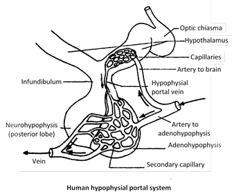
(1) Hypothalamo-hypophysial portal system : Present in higher vertebrates (amphibia, reptiles, birds and mammals). Blood from hypothalamus is collected by hypophysial portal vein which ends in anterior lobe of pituitary gland. The superior hypophysial artery which bring blood into circle of willis bifurcate outside the lobe; one branch supplies the lobe itself, but the other one supplies the hypothalamus. The vein that drain the blood from hypothalamus then runs into pars distalis and divide into capillaries. Thus this is a portal vein called hypothalamo-hypophysial portal vein.
Function : This portal system enables the releasing factors and inhibiting factors from hypothalamus to reach upto anterior pituitary.
(2) Hepatic portal system : Found in all chordates. In mammals, there is a single vein called hepatic portal vein, formed by the union of six main veins, which drain venous blood from different parts of alimentary canal (digestive system) into the liver. These veins are :
(i) Posterior or Inferior mesenteric vein : Collect blood from rectal wall and anal region. This vein possess maximum diluted blood. Posterior mesenteric made up of by joining of 4 small veins that is rectal vein, sigmoid vein, left colonic vein and it opens into the splenic vein.
(ii) Anterior or Superior mesenteric vein : Collect blood from wall of colon, caecum and small intestine. This vein possesses largest concentration of nutrients (glucose, amino-acid and vitamins). This vein formed by the joining of right colonic vein, ileocolic vein and appendicular vein.
(iii) Splenic vein : Collect blood from spleen and pancreas, splenic vein possess free haemoglobin in large amount.
(iv) Right gastric vein : Receives blood from stomach.
(v) Left gastric vein : Receives blood from stomach and pancreas.
(vi) Cystic vein : Receives blood from gall bladder.
Posterior mesenteric vein open into splenic vein and splenic, anterior mesenteric, right gastric fused to form hepatic portal vein, which leads blood in to the liver.
In amphibians (example - frog), hepatic portal system is formed of single hepatic portal vein and single anterior abdominal vein. The latter collects blood from leg region and drains it into the left lobe of liver.
Significance of hepatic portal system : The hepatic portal system has following significance.
(a) The blood which comes from the alimentary canal contains digested food like glucose and amino acids. The excess of glucose is converted into glycogen which is stored in the liver for later use. When an individual feels deficiency of food, the glycogen is converted into glucose and is transferred to the blood stream via hepatic veins.
(b) Harmful nitrogenous waste like ammonia is converted into urea which is later removed more...
It is a part of greater circulation which begins in the tissue fluid with lymphatic capillaries which are always terminally closed. This system terminates into venous system near heart. The main components of this system are :
(1) Lymph : Lymph can be defined as blood minus RBC's. In addition to the blood vascular system all vertebrate possess a lymphatic system. It is colourless or yellowish fluid present in the lymph vessels. It is a mobile connective tissue like blood and is formed by the filtration of blood. This process involves the diffusion of substances from blood capillaries into the interstitial space which is, thus, the primary site of lymph formation. Two forces bring about a steady filtration of plasma fluid into the tissue spaces : capillary pressure \[(30-35\,\,mm\text{ }Hg)\] and colloid osmotic pressure in tissue fluid (8 mm Hg). After absorption by veins, a small amount of \[C{{O}_{2}}\] and waste material still remains in the tissue fluid which is absorbed in the lymphatic capillaries as lymph. So, we can say that lymph is modified tissue fluid.
Differences between lymph and blood
The form, structure and function of heart exhibits much variation. The characteristics of heart of fishes, amphibians, reptiles, birds and mammals is presented in the following table.
Heart of vertebrates
S.No.
Class of vertebrates
Characteristics
Example
Diagram
1.
Pisces (= Branchial heart), Cyclostomata
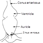
Thick, muscular, made of cardiac muscles, has two chambers (i) auricle and (ii) ventricle. The heart is called venous heart since it pumps deoxygenated blood to gills for oxygenation. This blood goes directly from gills to visceral organs (single circuit circulation). A sinus venosus and conus arteriosus is present. Lung fishes have only one auricles and one ventricle.
Labeo
Scoliodon
2.
Amphibians, Lung fish
Heart consists of :
(i) Two auricles
(ii) Undivided ventricle
(iii) Sinus venosus
(iv) Truncus arteriosus
(conus + proximal part of aorta) Right auricle receives blood from all the visceral organs (deoxygenated) via precaval and post caval. Pulmonary artery carries deoxygenated blood to more...
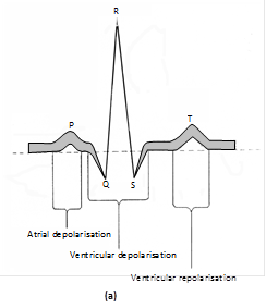
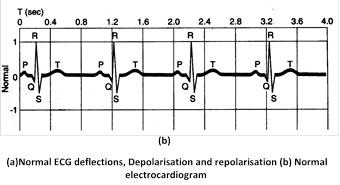
A graphic record of electrical events occuring during a cardiac cycle is called Electrocardiogram. The instrument used for recording the heart’s electrical variations is called Electrocardiograph in which the potential differences of heart muscles are recorded by a galvanometer. In ECG, there are 2 types of waves :
(1) Depolarisation waves : They represent the generation of the potential difference. These waves appear only when both electrodes of galvanometer are in different fields. When both the electrodes are in same field, there is no deflection and wave drops down to base line.
(2) Repolarisation waves : They appear when depolarisation is over and the muscle fibre is returning to its original polarity. When both electrodes are in same polarity (means 100% repolarisation and 100% depolarisation), there is no deflection.
A normal ECG has 5 deflection waves \[P,Q,R,S\] and T. Out of them \[P,R\] and T waves are above the base line and are called positive waves. The Q and S waves are below base line and are called negative waves. The part of the base line between any 2 deflections is called Interval.
P wave : Indicates impulse of contraction generated by S.A. node and its spread in atria causing atrial depolarisation. The interval PQ represents atrial contraction and takes 0.1 second.
QRS complex : Indicates spread of impulse of contraction from A.V node to the wall of ventricles through bundle of His and purkinje fibres causing ventricular depolarisation. This complex also represents repolarization of S.A. node.
The RS of QRS wave and ST interval show ventricular contraction (0.3 seconds). QRS is related to ventricular systole.
T wave : Indicates repolarisation during ventricular relaxation.
Any abnormality in the working of heart alters the wave pattern of ECG. Thus, ECG is of great diagnostic value in cardiac diseases. ECG also indicates the rate of heart beat
During the completion of one heart beat is called as cardiac cycle. Following events are repeated in a cyclic manner during each heart beat.
(1) Auricular systole : The atria contract due to wave of contraction stimulated by S.A. node contraction of auricles drives most of their blood into respective ventricles as the A.V. valves are open. There is no backflow of blood into the large veins as the contraction begins at the upper end and passes towards ventricles and moreover, the valves present at the opening of these veins close. Also, blood is already present in large veins which offers resistance to the blood that may return from the atria. At the end of a atrial systole, there starts the relaxation of auricles (auricular diastole) and contraction of ventricles (ventricular systole) simultaneously. Atrial systole takes 0.1 second while atrial diastole is of about 0.7 seconds.
(2) Ventricular systole : The ventricles begin to contract due to a wave of contraction stimulated by A.V. node. Due to ventricular systole, the pressure of blood in ventricles immediately rises above that in the auricles. With this pressure, the bicuspid and tricuspid valves close rapidly to prevent the backflow of blood. This closure of A.V. valves at the start of ventricular systole produces first heart sound called “Lubb” or Systolic sound. The semilunar valves are also close at this time. When the pressure of blood in the ventricles exceeds that in the great arteries, the semilunar valves open and blood enters into the great arteries. This marks the end of ventricular systole which takes about 0.3 seconds. Now the ventricles start relaxing (ventricular diastole which lasts for about 0.5 sec.)
(3) Joint diastole : The ventricles and auricles are in the diastolic phase simultaneously. As the ventricular diastole progresses, the pressure in the ventricles falls below that in the great arteries. So, to prevent backflow of blood from great arteries into ventricles, the semilunar valves close rapidly. This rapid closure of semilunar valves at the beginning of ventricular diastole produces second heart sound “Dup” or diastolic sound.
The quality of heart sounds indicates the state of the heart valves. Defective or damaged heart valves lead to the backflow of blood either from ventricles to auricles or from aortae to ventricles. Such defects are detectable as abnormal hissing sound called “Murmur”. Defective valves may be replaced or repaired surgically. Syphilis and Rheumatic fever cause Murmur. The instrument used to magnify and record the heart sound is called Phonocardiogram.
During joint diastole, blood from great veins and coronary sinus flows into the atria and some blood also passes from auricles into the respective relaxing ventricles due to less pressure in ventricles. This phase takes only 0.4 seconds and is also called as blood receiving period of heart. Thus a cardiac cycle is completed in 0.8 seconds.
Cardiac output : Volume of blood pumped from heart (left ventricle) into the systemic aorta in one minute is called cardiac output. It is also called minute volume. It more...
The study of blood vessels is called Angiology. The blood vessels are of following types :
(1) Arteries : Thick walled, carrying oxygenated blood (deoxygenated in pulmonary artery) from heart to various parts of body. These blood vessels are grouped as Aorta which branches to form arteries which further divides into thinner branches called arterioles inside the organ. Average diameter of arteriole is \[120\,\,\mu m\] the arterioles further divide into smaller vessels called meta-arterioles \[(70\,\,\mu m)\] which divide into capillaries. At the beginning of capillary, the arterioles posses circular muscles called precapillary sphincter which regulates flow of blood into the capillaries which is called vasomotion. Smooth muscles of arteries innervated by sympathetic fibers, their stimulation control vasoconstriction and vasodilation. Smooth muscles of arteries and arterioles also limit bleeding from wounds by producing vascular spasm during cut. Arteries two types.
(i) Conducting or elastic arteries
(ii) Distributing or muscular arteries.
Elastic or conducting arteries receive blood from heart and do not provide it to any organ rather they provide blood to other atreries and are pressure reservoirs of blood.
Muscleless end of meta-arteriole is called thoroughfare channel or preferential channel.
The largest artery is dorsal / abdominal aorta (systemic aorta).
Anastomosis : If more than one arteries are supplying to one organ then branches of these arteries unite to form a network called Anastomosis. It provides many collateral or alternate pathways of blood supply. So, if there is blocking of any artery, it will not lead to necrosis.
(2) Capillaries : Smallest blood vessels, discovered by Marcello Malpighi (also layered nucleated squamous epithelial cells called endothelium resting on a basement membrane. Diameter of capillary is about 8m. These are also called as exchange vessels as they are the site of exchange of material between blood and tissue because of least barrier in them. The capillaries can be grouped into two categories :
(i) Arteriolar capillary : Which supplies nutrition, respiratory gases etc. to the body cells.
(ii) Veinular capillaries : Which collect the metabolic wastes from the body cells.
Capillaries possess about 7% of total body blood and are present near almost all cells of body in the intercellular spaces. The tissues which are devoid of intercellular spaces are also devoid of capillary. They are called avascular tissues.
Capillaries are surrounded by cells of connective tissue called pericapillary cells. Some of these cells are contractile and phagocytic in nature and are called Rouget cells or pericytes.
Continuous capillaries are without fenestra/aperture, hence are less permeable. These are present in organs such as lungs, muscles, connective tissues and brain tissues.
Fenestrated capillaries possess apertures/fenestra and are found in those organs where there is maximum need of permeability such as endocrine glands, intestinal villi, cavities of brain, kidney, ciliary body of eye.
Sinusoids are irregularly dilated capillaries found in organs where there is decrease in flow rate such as liver, spleen, bone marrow, parathyroid, pituitary gland. In liver, sinusoids are branches of venules and open into venules while more...
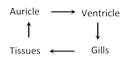
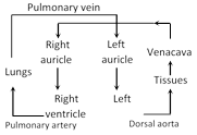
Blood circulation was discovered by William harvey. In case of vertebrates, blood circulation is of closed type, which can be grouped into two categories :
(1) Single circulation (2) Double circulation
Differences between single and double circulation
S.N.
Single circulation
Double circulation
1.
Blood flows only once through the heart in a complete cycle.
Blood flows in two circuit pulmonary and systemic.
2.
Heart pumps only deoxygenated blood, hence called Venous Heart.
Heart pumps both deoxygenated and oxygenated blood to lungs and body respectively, hence called arteriovenous heart.
3.
Blood is oxygenated in gills.
Blood is oxygenated in lungs.
4.
Less efficient as gill capillaries slow down the blood flow. more...
 (ii) Pronator and Supinator : The contraction of a pronator rotates the forearm to turn the palm downward or backward. Supinator is antagonist of pronator. A supinator contracts to rotate the forearm and thus to make palm face upward or forward.
 (iii) Abductor and Adductor : An abductor contracts to draw a bone away from the body midline. Muscle that brings the limb away from midline is called abductor. An adductor draws a bone towards the body midline. Muscles that brings the limb towards midline is called adductor. Abductor muscle is antagonist of adductor muscle. Abduction is elevation and adduction is depression.
 (2) Hepatic portal system : Found in all chordates. In mammals, there is a single vein called hepatic portal vein, formed by the union of six main veins, which drain venous blood from different parts of alimentary canal (digestive system) into the liver. These veins are :
(i) Posterior or Inferior mesenteric vein : Collect blood from rectal wall and anal region. This vein possess maximum diluted blood. Posterior mesenteric made up of by joining of 4 small veins that is rectal vein, sigmoid vein, left colonic vein and it opens into the splenic vein.
(ii) Anterior or Superior mesenteric vein : Collect blood from wall of colon, caecum and small intestine. This vein possesses largest concentration of nutrients (glucose, amino-acid and vitamins). This vein formed by the joining of right colonic vein, ileocolic vein and appendicular vein.
(iii) Splenic vein : Collect blood from spleen and pancreas, splenic vein possess free haemoglobin in large amount.
(iv) Right gastric vein : Receives blood from stomach.
(v) Left gastric vein : Receives blood from stomach and pancreas.
(vi) Cystic vein : Receives blood from gall bladder.
Posterior mesenteric vein open into splenic vein and splenic, anterior mesenteric, right gastric fused to form hepatic portal vein, which leads blood in to the liver.
In amphibians (example - frog), hepatic portal system is formed of single hepatic portal vein and single anterior abdominal vein. The latter collects blood from leg region and drains it into the left lobe of liver.
Significance of hepatic portal system : The hepatic portal system has following significance.
(a) The blood which comes from the alimentary canal contains digested food like glucose and amino acids. The excess of glucose is converted into glycogen which is stored in the liver for later use. When an individual feels deficiency of food, the glycogen is converted into glucose and is transferred to the blood stream via hepatic veins.
(b) Harmful nitrogenous waste like ammonia is converted into urea which is later removed more...
(2) Hepatic portal system : Found in all chordates. In mammals, there is a single vein called hepatic portal vein, formed by the union of six main veins, which drain venous blood from different parts of alimentary canal (digestive system) into the liver. These veins are :
(i) Posterior or Inferior mesenteric vein : Collect blood from rectal wall and anal region. This vein possess maximum diluted blood. Posterior mesenteric made up of by joining of 4 small veins that is rectal vein, sigmoid vein, left colonic vein and it opens into the splenic vein.
(ii) Anterior or Superior mesenteric vein : Collect blood from wall of colon, caecum and small intestine. This vein possesses largest concentration of nutrients (glucose, amino-acid and vitamins). This vein formed by the joining of right colonic vein, ileocolic vein and appendicular vein.
(iii) Splenic vein : Collect blood from spleen and pancreas, splenic vein possess free haemoglobin in large amount.
(iv) Right gastric vein : Receives blood from stomach.
(v) Left gastric vein : Receives blood from stomach and pancreas.
(vi) Cystic vein : Receives blood from gall bladder.
Posterior mesenteric vein open into splenic vein and splenic, anterior mesenteric, right gastric fused to form hepatic portal vein, which leads blood in to the liver.
In amphibians (example - frog), hepatic portal system is formed of single hepatic portal vein and single anterior abdominal vein. The latter collects blood from leg region and drains it into the left lobe of liver.
Significance of hepatic portal system : The hepatic portal system has following significance.
(a) The blood which comes from the alimentary canal contains digested food like glucose and amino acids. The excess of glucose is converted into glycogen which is stored in the liver for later use. When an individual feels deficiency of food, the glycogen is converted into glucose and is transferred to the blood stream via hepatic veins.
(b) Harmful nitrogenous waste like ammonia is converted into urea which is later removed more...