Current Affairs 11th Class

It is the process by which a mature cell divides and forms two nearly equal daughter cells which resemble the parental cell in a number of characters. In unicellular organisms, cell division is the means of reproduction by which the mother cell produces two or more new cells. In multicellular organism also, new individual develop from a single cell. Cell division is central to life of all cell and is essential for the perpetuation of the species. Discovery : Prevost and Dumans (1824) first to study cell division during the cleavage of zygote of frog. Nagelli (1846) first to propose that new cells are formed by the division of pre-existing cells. Rudolf Virchow (1859) proposed “omnis cellula e cellula” and “cell lineage theory”. A cell divides when it has grown to a certain maximum size which disturb the karyoplasmic index (KI)/Nucleoplasmic ratio (NP)/Kernplasm connection. Cell cycle : Howard and Pelc (1953) first time described it. The sequence of events which occur during cell growth and cell division are collectively called cell cycle. Cell cycle completes in two steps: (1) Interphase,                  (2) M-phase/Dividing phase (1) Interphase : It is the period between the end of one cell division to the beginning of next cell division. It is also called resting phase or not dividing phase. But, it is actually highly metabolic active phase, in which cell prepares itself for next cell division. In case of human beings it will take approx 25 hours. Interphase is completed in to three successive stages. G1 phase/Post mitotic/Pre-DNA synthetic phase/gap Ist : In which following events take place. (i) Intensive cellular synthesis. (ii) Synthesis of rRNA, mRNA ribosomes and proteins. (iii) Metabolic rate is high. (iv) Cells become differentiated. (v) Synthesis of enzymes and ATP storage. (vi) Cell size increases. (vii) Decision for a division in a cell occurs. (viii) Substances of G stimulates the onset of next S – phase. (ix) Synthesis of NHC protein, carbohydrates, proteins, lipids. (x) Longest and most variable phase. (xi) Synthesis of enzyme, amino acids, nucleotides etc. but there is no change in DNA amount. S-phase/Synthetic phase (i) DNA replicates and its amount becomes double \[(2C-4C).\] (ii) Synthesis of histone proteins and NHC (non-histone chromosomal proteins). (iii) Euchromatin replicates earlier than heterochromatin. (iv) Each chromosome has 2 chromatids. G2-phase/Pre mitotic/Post synthetic phase/gap-IInd (i) Mitotic spindle protein (tubulin) synthesis begins. (ii) Chromosome condensation factor appears. (iii) Synthesis of 3 types of RNA, NHC proteins, and ATP mole. (iv) Duplication of mitochondria, plastids and other cellular macromolecular complements. (v) Damaged DNA repair occur. (2) M-phase/Dividing phase/Mitotic phase : It is divided in to two phases, karyokinesis and cytokinesis.      
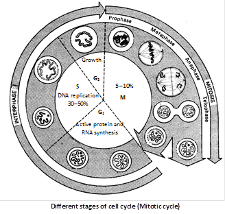
Duration of cell cycle : Time period for \[{{G}_{1}},S,{{G}_{2}}\] and M-phase is species specific under specific environmental conditions. e.g., 20 minutes for bacterial cell, 8-10 hours for intestinal epithelial cell, and onion root tip cells may take 20 hours. \[{{\mathbf{G}}_{\mathbf{0}}}\mathbf{-}\]phase (Lajtha, 1963) : The cells, which are not to divide further, do more...

Within the cytoplasm of a cell there occur many different kinds of non-living structures which are called inclusions or ergastic / Deutoplasmic substances. (1) Vacuoles : The vacuole in plants was discovered by Spallanzani. It is a non-living reservoir, bounded by a differentially or selectively permeable membrane, the tonoplast. The vacuole is filled with cell sap or tonoplasm. They contain water, minerals and anthocyanin pigments. Some protozoans have contractile vacuoles which enlarge by accumulation of fluid or collapse by expelling them from the cell. The vacuoles may be sap vacuoles, contractile vacuoles or gas vacuoles (pseudo vacuoles). Function of vacuoles : Vacuole maintains osmotic relation of cell which is helpful in absorption of water. Turgidity and flaccid stages of a cell are due to the concentrations of sap in the vacuole. (2) Reserve food material The reserve food material may be classified as follows : (i) Carbohydrates : Non-nitrogenous, soluble or non- soluble important reserve food material. Starch cellulose and glycogen are all insoluble. (a) Starch : Found in plants in the form of minute solid grains. Starch grains are of two types : Assimilation starch : It is formed as a result of photosynthesis of chloroplasts. Reserve starch : Thick layers are deposited around an organic centre called hilum. (b) Glycogen : Glycogen or animal starch occurs only in colourless plants like fungi. (c) Inulin : It is a complex type of polysaccharide, soluble and found dissolved in cell sap of roots of Dahlia, Jaruslem, Artichoke, Dandelion and members of compositae. (d) Sugars : A number of sugars are found in solution of cell sap. These include glucose, fructose, sucrose, etc. (e) Cellulose : Chemical formula is \[{{({{C}_{6}}{{H}_{10}}{{O}_{5}})}_{n}}.\]The cell wall is made up of cellulose. It is insoluble in water. (ii) Fats and Oils : These are important reserve food material. These are always decomposed into glycerol and fatty acids by enzymatic action. Fat is usually abundant in cotyledons than in the endosperm. e.g., flax seed produce linseed oil, castor produce castor oil, cotton seeds produce cottonseed oil, etc. (iii) Proteins and Amides (Aleurone grains) : Storage organ usually contain protein in the form of crystalline bodies known as crystalloids (potato). Proteins may be in the form of aleurone grains as in pea, maize, castor, wheat, etc. (3) Excretory Products : The organic waste products of plants are by-product of metabolism. They are classified as : (i) Resins : They are believed to be aromatic compounds consisting of carbon, hydrogen and oxygen and are acidic in nature. Sometimes they are found in combination with gums and are called gum resin. e.g., Asafoetida (heeng). (ii) Tannins : They are complex nitrogenous compounds of acid nature having an astringent taste. Presence of tannin in plants makes its wood hard durable and germ proof. (iii) Alkaloids : These are organic, basic, nitrogenous substance. They occur in combination with organic acids and most of them are poisonous. From plants, cocaine, hyoscine, morphine, nicotine, quinine, atropine, strychnine and daturine etc. are extracted. (iv) Glucosides more...

The nucleus also called director of the cell. It is the most important part of the cell which directs and controls all the cellular function. Discovery : The nucleus was first observed by Robert Brown (1831), in orchid root cells. Nucleus plays determinative (in heredity) role in cell and organism, that was experimentally demonstrated by Hammerling (1934) in surgical experiments with green marine unicelled algae Acetabularia. Occurence : A true nucleus with definite nuclear membrane and linear chromosome, is present in all the eukaryotes except mature mammalian RBCs, sieve tube cell of phloem, tracheids and vessels of xylem. The prokaryotes have an incipient nucleus, called nucleoid or prokaryon or genophore or false nucleus. Number : Usually there is a single nucleus per cell i.e., mononucleate condition, e.g., Acetabularia. (1) Anucleate (without nucleus) : RBCs of mammals, phloem sieve tube, trachids and vessels of xylam. (2) Binucleate : e.g., Ciliate, Protozoans like Paramecium. (3) Polynucleate : e.g., fungal hyphae of Rhizopus, Vaucheria. Polynucleate condition may be because of fusion of a number of cells. i.e., syncytium, coconut endosperm or by free nuclear divisions without cytokinesis i.e., coenocyte. Shape : It varies widely, generally spherical e.g., cuboidal germ cells, oval e.g., columnar cells of intestine, bean shaped  in paramecium, horse-shoe shaped in Vorticella, bilobed, e.g., WBCs (acidophils), 3 lobed e.g., basophil, multilobed e.g., neutrophils, long and beaded form (moniliform) e.g., stentor and branched in silk spinning cells of platy phalyx insect larva. Size : The size of nucleus is variable i.e., \[530\mu .\] In metabolically active cells size of the nucleus is larger than metabolically inactive cells. Chemical composition Proteins \[=80%,\text{ }DNA=12%,\text{ }RNA=5%,\text{ }Lipids=3%\] Enzymes like polymerases are abundantly present and help in synthesis of DNA and RNA. Ultrastructure : The nucleus is composed of following structure. (1) Nuclear membrane : It is also called nuclear envelope or nucleolemma or karyotheca, was first discovered by Erclab (1845). Structure : It is a bilayered envelope. Each membrane is about 60-90Å thick lipoproteinous and trilaminar. Outer membrane, called ectokaryotheca (with ribosome) and inner membrane is called endokaryotheca (without ribosome). Two membranes are separated by a fluid-filled intermembranous perinuclear space (about \[100-300{AA}\]). Nuclear membrane is porous and has \[1,000-10,000\] octagonal nuclear pores. Each nuclear pore is about \[400-1,000\text{ }{AA}\] in diameter (average size is \[800\text{ }{AA}\]). Callan and Tamlin (1950) first to observe nuclear pore in nuclear membrane. The nuclear pares are enclosed by circular structure are called annuli. The pore and annuli together are called pore complex or pore basket.     
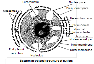
Origin : It is formed by the fusion of ER elements during the telophase of cell division. Functions (i) It regulates the nucleo-cytoplasmic interactions. (ii) It allows the passage of inorganic ions, small organic molecules, ribosomal subunits, RNAs and proteins through nuclear pores. (iii) It maintains the shape of the nucleus. (2) The nucleolus (Little nucleus plasmosome) : It was first observed by Fontana (1781) in the skin cells of an eel. Bowman (1840) more...

Chromosome were discover by Hofmeister (1848) in filament of pollen mother cells of tradescantia (Rhoeodiscolour) studied by strasburger (1875) and given the persent name by Waldeyer (1888). During interphase, chromatin threads are present in the form of a network called chromatin reticulum. At the time of cell division, these thread like structures of chromatin become visible as independent structures, called chromosomes. The haploid set of chromosomes is define as genome. Structure : Each chromosome consists of two coiled filaments throughout its length called chromonemata by Vejdovsky. These have bead like structures called chromomeres which bear genes. Chromatid is a half chromosome or daughter chromosome. The two chromatids are connected at the centromere or primary constriction. Primary constriction (centromere) and secondary constriction gives rise to satellite. The secondary constriction consists of genes which code for ribosomal RNA and nucleolus hence it is called as “nucleolar organizer region”. Chromosomes having satellite are called SAT chromosomes. The ends of chromosomes are called “telomeres” (which do not unite with any other structure). In 1928 Emile Heitz developed a technique for stainning of chromosomes. Staining property of chromosomes is called as heteropycnosis. Chromosomes can be stained with basic dye like janus green there are two types of regions are seen :– (1) Heterochromatin : It is formed of thick regions which are more darkly stained than others areas. It is with condensed RNA which is transcriptionally inactive and late replicating. It generally lies near the nuclear lamina. It is of two type : (i) Constitutive hetrochromative : Occurs in all cells in all stages. e.g., Centromere. (ii) Facultative hetrochromative : Formed by inactivation of some gene in some cell in some stages. e.g., Barr body.  (2) Euchromatin : It is true chromatin and is formed of thin, less darkly stained  areas. It is with loose DNA which is transcriptionally active and early replicating. Chemical chomposition : DNA - 40%. Histone – 50%. Other (acid) Proteins – 8.5%. RNA – 1.5%. Traces of lipids, Ca, Mg and Fe. Histone are low molecular weight basic proteins which occur alongwith DNA in ratio. Nonhistone chromosomal or NHC proteins are of three types– structural, enzymatic and regulatory. Structural NHC proteins form the core or axis of the chromosome. They are also called scaffold proteins.

Chatton gave the term prokaryote and eukaryote. Depending upon the nature of nucleus cells are classified. Incipient nucleus is present in prokaryotes, where as in eukaryotes well organised nucleus is present.   Differences between prokaryotic and eukaryotic cell more...
The ribosomes are smallest known electron microscopic without membrane, ribonucleo–protein particles attached either on RER or floating freely in the cytoplasm and are the sites of protein synthesis.  Discovery : In 1943 Claude observed some basophilic bodies and named them as microsome. Palade (1955) coined the term ribosome (form animal cell). Ribosomes in nucleoplasm were observed by Tsao and Sato (1959). First isolated by Tissieres and Watson (1958) from E. coli. Ribosomes found in groups are termed as polyribosomes or ergosomes (Rich and Warner 1963 observed first time polyribosomes). Occurrence : In prokaryotes ribosomes are found only in free form in the cytoplasm. While in the eukaryotes the ribosomes are found in two forms in the cytoplasm, free form and bind form (bound on RER and outer nuclear membrane). These are also reported inside some cell organelles like mitochondria and plastids respectively called mitoribosomes and plastidoribosomes. Types of ribosomes (1) 70S ribosomes : Found in prokaryotes, mitochondria and plastid of eukaryotes. (2) 80S ribosomes : Found in cytoplasm of eukaryotes. (3) 77S, 60S and 55S ribosomes : Levine and Goodenough (1874) observed 77S ribosomes in fungal mitochondria 60S ribosomes in animal mitochondria and 55S in mammalian  mitochondria. Structure : Each ribosome is formed of two unequal subunits, which join only at the time of protein synthesis. In 70S and 80S ribosomes, 50S and 30S, 60S and 40S are larger and smaller subunits respectively. Larger subunits is dome shaped and attached to ER by glycoproteins called “ribophorins”.     Smaller subunit is oval shaped and fits as a cap on flat side of larger subunit. Ribosomes are attached to ER through hydrophobic interactions. Chemical composition : Ribosomes are chemically composed of rRNA and proteins Ribonucleo-Protein (RNP). 70S ribosomes has 60-65% rRNA and \[35-40%\] proteins (ratio is 1.5:1). rRNAs are of three types : 23S type and 5S type rRNAs in 50S and 16S type rRNA in 30S sub-units. 80S ribosome has 45% rRNA and 55% proteins (ratio is about 1 : 1). r-RNA are of four types : 28S, 5S and 5.8S types of rRNAs in 60S and 18S type rRNA in 40S sub-units. A \[1\times {{10}^{-3}}\,(0.001M)\] molar concentration of \[M{{g}^{++}}\] is needed for the structural cohesion of ribosomes i.e., for holding the two subunits together. If this concentration is increased by ten folds, two ribosomes unite to form a dimer. By decreasing the \[M{{g}^{++}}\] conc. to normal, the dimer breaks into monomers (single ribosomes). Biogenesis of ribosome (1) In eukaryotes the ribosomal RNAs like 18S, 5.8S and 28S are synthesized by nucleolus and 5S RNA out of the nucleus.   (2) In prokaryotes both rRNA and its protein are synthesized as well as assembled by cytoplasm. Polyribosomes or Polysomes : When many ribosomes (generally \[68\]) are attached at some mRNA strand. It is called polysome. The distance between adjacent ribosomes is of 90 nucleotides. These are functional unit of protein synthesis. Functions (1) Ribosomes are also called protein factories of the cell or
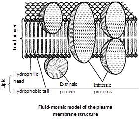
 more...

Protoplasm is a complex, granular, elastic, viscous and colourless substance. It is selectively or differentially permeable. It is considered as “Polyphasic colloidal system”. Discoveries (1) J. Huxley defined it as “physical basis of life”. (2) Dujardin (1835) discovered it and called them “sarcode”. (3) Purkinje (1837) renamed it as “Protoplasm”. (4) Hugo Von Mohl (1844) gave the significance of it. (5) Max Schultz (1861) gave the protoplasmic theory for plants. (6) Fischer (1894) and Hardy (1899) showed its colloidal nature. (7) Altman (1893) suggested protoplasm as granular.   Chemically Composition
Prokaryotic cell Eukaryotic cell
It is a single membrane system. It is a double membrane system.
Cell wall surrounds the plasma membrane. Cell wall surrounds the plasma membrane in some protists, most fungi and all plant cell. Animal cell lacks it.
Cell wall is composed of peptidoglycans. Strengthening material is murein. It is composed of polysaccharide. Strengthening material is chitin in fungi and cellulose in others plants.
Cell membrane bears respiratory enzymes. It lacks respiratory enzymes.
Cytoplasm lacks cell organelles e.g., Mitochondria, ER, Golgi body etc. Cytoplasm contains various cell organelles.
Ribosomes are only 70 S type. Ribosomes are both 80 S and 70 S type.
There are no streaming movements of cytoplasm. Cytoplasm show streaming movements.
Water 75 - 85% Carbon 20%
Proteins 10 - 25% Oxygen 62%
Lipids 2 - 3% Hydrogen 10%
Inorganic Materials 1% Nitrogen 3%
Trace elements 5% (Ca, P, Cl, more...
Plastids are semiautonomous organelles having DNA, RNA, Ribosomes and double membrane envelope. These are largest cell organelles in plant cell. History (1) Haeckel (1865) discovered plastid, but the term was first time used by Schimper (1883). (2) A well organised system of grana and stroma in plastid of normal barley plant was reported by de Von Wettstein. (3) Park and Biggins (1964) gave the concept of quantasomes. (4) The term chlorophyll was given by Pelletier and Caventou, and structural details were given by Willstatter and Stall. (5) The term thylakoid was given by Menke (1962). (6) Fine structure was given by Mayer. (7) Ris and Plaut (1962) reported DNA in chloroplast and was called plastidome. Types of plastids : According to Schimper, Plastids are of 3 types: Leucoplasts, Chromoplasts and Chloroplasts. Leucoplasts : They are colourless plastids which generally occur near the nucleus in nongreen cells and possess internal lamellae. Grana and photosynthetic pigments are absent. They mainly store food materials and occur in the cells not exposed to sunlight e.g., seeds, underground stems, roots, tubers, rhizomes etc. These are of three types. (1) Amyloplast : Synthesize and store starch grains. e.g., potato tubers, wheat and rice grains. (2) Elaioplast (Lipidoplast, Oleoplast) : They store lipids and oils e.g., castor endosperm, tube rose, etc. (3) Aleuroplast (Proteinoplast) : Store proteins e.g., aleurone cells of maize grains. Chromoplasts : Coloured plastids other than green are kown as chromoplasts. These are present in petals and fruits. These also carry on photosynthesis. These may arise from the chloroplasts due to replacement of chlorophyll by other pigments. Green tomatoes and chillies turn red on ripening because of replacement of chlorophyll molecule in chloroplasts by the red pigment lycopene in tomato and capsanthin in chillies. Thus, chloroplasts are changed into chromatoplast. All colours (except green) are produced by flavins, flavenoids and cyanin. Cyanin pigment is of two types one is anthocyanin (blue) and another is erythrocyanin (red). Anthocyanin are water soluble pigments and found in cell sap of vacoule. Chloroplast : Discovered by Sachs and named by Schimper. They are greenish plastids which possess photosynthetic pigments. Number : It is variable. Number of chloroplast is 1 in Spirogyra indica, 2 in Zygnema, 16 in S.rectospora, up to 100 in mesophyll cells. The minimum number of one chloroplast per cell is found in Ulothrix and species of Chlamydomonas. Shape : They have various shapes  
Shape Example
Cup shaped more...
Every living cell is externally covered by a thin transparent electron microscopic, elastic regenerative and selective permeable membrane called plasma membrane. It is quasifluid in nature. Membranes also occur inside the cells. They are collectively called biomembranes. The term cell membrane was given by C. Nageli and C. Cramer (1855) for outer membrane covering of the portoplast. It was replaced by the term plasmalemma by Plower (1931). Chemical composition : Proteins lipoprotein (Lipid +Protein) are the major component forming 60% of the plasma membrane. Proteins provide mechanical strength and responsible for transportation of different substances. Proteins also act as enzyme. Lipids account may 28%-79% depending upon the type of cell and organism involved (in humans, myelin 79%). The lipids of plasma membrane are of three types namely phospholipids, glycolipids and sterols. The sterol found in the membrane may be cholesterol (Animals), phytosterol (Plants) or ergosterol (Microorganisms). Carbohydrates form 2%–10%. Oligosaccharides are the main carbohydrates present in plasma membrane. The carbohydrates of plasma membrane are covalently linked to both lipid and protein components. Ultrastructure : Under electron microscope the plasma membrane appears three layered, i.e., trilaminar or tripartite. One optically light layer is of lipid and on both sides two optically dense protein layers are present. Molecular structure and different models : Several models have been proposed to explain the structure and function of the plasma membrane. (1) Overton’s model : It suggests that the plasma membrane is composed of a thin lipid single layer. (2) Sandwitch model : It was proposed by Davson and Danielli (1935). According to this model the light biomolecular lipid layer is sandwitched between two dense protein layers (globular a type protein). This model was also said to be unit membrane hypothesis. (3) Robertson’s unit membrane model : It states that all cytoplasmic membranes have a similar structure of three layers with and electron transparent phospholipid bilayer being sandwitched between two electron dense layer of proteins (extended or \[\beta \]  type protein). Its thickness is about 75 Å with a central lipid layer of \[35\text{ }{AA}\] thick and two peripheral protein layers of \[\text{20 }{AA}\] thick. (4) Fluid mosaic model : The most important and widely accepted latest model for plasma membrane was given by Singer and Nicolson in 1972. According to them it is “protein iceberg in a sea of lipids.” According to this model, the cell membrane consists of a highly viscous fluid matrix of two layers of phospholipid molecules. Protein molecules occur as separate particles asymmetrical arranged in a mosaic pattern. Some of these are loosely bound at the polar surfaces of lipid layers, called peripheral or extrinsic proteins. Others penetrate deeply into the lipid layer called integral or intrinsic proteins. Some of the integral proteins penetrate through the phospholipid layers and project on both the surface. These are called trans membrane or tunnel proteins (glycophorins). Singly or in groups, they function as channels for passage of water ions and other solutes.     The more...

(1) Sphaerosomes Discovery : These were first observed by Hanstein (1880) but discovered by Perner (1953). Term sphaerosomes was given by Dangeard. Occurrence : These are found in all the plant cells which involves in the synthesis and storage of lipids i.e., endosperm and cotyledon of oil seeds. Shape, size and structure : These are spherical or oval in shape about \[0.5-2.5\,\mu \,m\]in diameter. They contain hydrolytic enzymes like protease, ribonuclease, phosphatase, esterase etc. They are bounded by a single unit membrane. Function : The main function of sphaerosomes is to help in lipid metabolism. These are also known as plant lysosomes. (2) Peroxisomes (Uricosomes) Discovery : These were first discovered by J. Rhodin (1954) in the cells of mouse kidney and were called microbodies. De Duve (1965) isolated certain sac like organelles from various types of animals and plants. These were called peroxisomes because these contain peroxide producing enzymes (oxidases) and peroxide destroying enzymes (catalases). Occurrence : These are found in photosynthetic cells of plants. In animals peroxisomes are found in vertebrates (cells of liver, kidney), brain, small intestine, testis and adrenal cortex), invertebrates and protozoans e.g., Paramecium. Shape, size and structure : These are spherical in shape, about \[1.5\,\mu \,m\] in size. They are bounded by a single unit membrane. Their membrane is permeable to amino acids, uric acids, etc. They contain four enzymes of \[{{H}_{2}}{{O}_{2}}\] metabolism. The enzymes urate oxidase, d-amino oxidase, a-hydroxy acid oxidase produce \[{{H}_{2}}{{O}_{2}}\] whereas the catalases plays a significant protective role by degrading H2O2 because \[{{H}_{2}}{{O}_{2}}\] is toxic for cells. Function : These are involved in the formation  and degrading of \[{{H}_{2}}{{O}_{2}}\]. Plant peroxisomes are also involved in photorespiration. (3) Glyoxysomes Discovery : These were discovered by Beevers in 1961 and Briedenbach in 1967. Occurrence : These are found in fungi, some protists and germinating fatty seeds where insoluble lipid food reserves must be turned into soluble sugars. Absent in animal cell. Shape, size and structure : These are spherical in shape, about \[0.5-1\,\mu \,m\]in size, they contain enzymes of metabolism of glycolic acid via glyoxylate cycle and bounded by a unit membrane. These are also contain enzymes for \[\beta -\]oxidation of fatty acids. Produced acetyl CoA. The better is metabolised in glyoxlate cycle to produced carbohydrates. Functions : The main function of glyoxysomes is conversion of fats into carbohydrates. (4) Lomasomes : These are sac like structures found between cell wall and plasmalemma in the haustoria of fungal hyphae. These were first discovered by Bowen and Berlin. Webster called them border bodies.


You need to login to perform this action.
You will be redirected in 3 sec spinner