 Class 1. Chondrichthyes (The Cartilaginous Fishes)
(Gk. chondros = cartilage; ichthys = fish)
General characters.
(1) Mostly marine and predaceous.
(2) Body fusiform or spindle shaped.
(3) Fins both median and paired, all supported by fin rays. Pelvic fins bear claspers in male. Tail heterocercal.
(4) Skin tough containing minute placoid scales and mucous glands.
(5) Endoskeleton entirely cartilaginous, without true bones. Notochord persistent. Vertebrae complete and separate. Pectoral and pelvic girdles present.
(6) Mouth ventral. Jaws present. Teeth are modified placoid scales. Stomach J-shaped. Intestine with spiral valve.
(7) Respiration by 5 to 7 pairs of gills. Gill-slits separate and uncovered (except, chimaeras). Operculum absent. No air bladder and lungs.
(8) Heart 2–chambered (1 auricle and 1 ventricle). Sinus venosus and conus arteriosus present. Both renal and portal systems present. Temperature variable (poikilothermous or cold blooded or ectothermal animal.
(9) Kidneys mesonephric or opisthonephric. Excretion ureotelic. Cloaca present.
(10) Brain with large olfactory lobes and cerebellum. Cranial nerves 10 pairs.
(11) Olfactory sacs do not open into pharynx. Membranous labyrinth with 3 semicircular canals. Lateral line system present.
(12) Sexes separate. Gonads paired. Gonoducts open into cloaca. Fertilization internal. Oviparous or ovoviviparous. Eggs large, yolky. Cleavage meroblastic. Development direct, without metamorphosis.
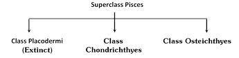
Classification of Chondrichthyes
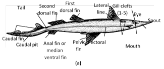
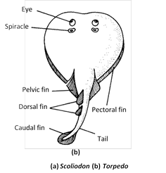
(a) Subclass I. Selachii : (Gk., selachos, a shark)
(1) Multiple gill slits on either side protected by individual skin flaps.
(2) A spiracle behind each eye.
(3) Cloaca present.
Examples : True sharks. Dogfishes (Scoliodon, Chiloscyllium, Mustelus, Carcharinus), spiny dogfish (Squalus) seven gilled shark (Heptanchus), Stegostoma, Sphyrna, Rhineodon. Skates and rays. Skate (Raja) Trygon, Torpedo, Myliobatis, Rhinobatus, Pristis.
• Zebra shark (Stegostoma) is the most beautiful fish in the sea.
Class 1. Chondrichthyes (The Cartilaginous Fishes)
(Gk. chondros = cartilage; ichthys = fish)
General characters.
(1) Mostly marine and predaceous.
(2) Body fusiform or spindle shaped.
(3) Fins both median and paired, all supported by fin rays. Pelvic fins bear claspers in male. Tail heterocercal.
(4) Skin tough containing minute placoid scales and mucous glands.
(5) Endoskeleton entirely cartilaginous, without true bones. Notochord persistent. Vertebrae complete and separate. Pectoral and pelvic girdles present.
(6) Mouth ventral. Jaws present. Teeth are modified placoid scales. Stomach J-shaped. Intestine with spiral valve.
(7) Respiration by 5 to 7 pairs of gills. Gill-slits separate and uncovered (except, chimaeras). Operculum absent. No air bladder and lungs.
(8) Heart 2–chambered (1 auricle and 1 ventricle). Sinus venosus and conus arteriosus present. Both renal and portal systems present. Temperature variable (poikilothermous or cold blooded or ectothermal animal.
(9) Kidneys mesonephric or opisthonephric. Excretion ureotelic. Cloaca present.
(10) Brain with large olfactory lobes and cerebellum. Cranial nerves 10 pairs.
(11) Olfactory sacs do not open into pharynx. Membranous labyrinth with 3 semicircular canals. Lateral line system present.
(12) Sexes separate. Gonads paired. Gonoducts open into cloaca. Fertilization internal. Oviparous or ovoviviparous. Eggs large, yolky. Cleavage meroblastic. Development direct, without metamorphosis.
Classification of Chondrichthyes
(a) Subclass I. Selachii : (Gk., selachos, a shark)
(1) Multiple gill slits on either side protected by individual skin flaps.
(2) A spiracle behind each eye.
(3) Cloaca present.
Examples : True sharks. Dogfishes (Scoliodon, Chiloscyllium, Mustelus, Carcharinus), spiny dogfish (Squalus) seven gilled shark (Heptanchus), Stegostoma, Sphyrna, Rhineodon. Skates and rays. Skate (Raja) Trygon, Torpedo, Myliobatis, Rhinobatus, Pristis.
• Zebra shark (Stegostoma) is the most beautiful fish in the sea.
 (b) Subclass II. Holocephali : (Gk., holos, entire + kephale, head)
(1) Single gill opening on either side covered by a fleshy operculum.
(2) No spiracles, cloaca and scales.
(3) Jaws with tooth plates.
(4) Single nasal opening.
(5) Lateral line system with open groove.
Examples : Hydrolagus (= Chimaera).
Class 2. Osteichthyes–(The Bony fishes)
(Gk. osteon = bone; ichtyes = fish)
General Characters
(1) Inhabit all sorts of water-fresh, brackish or salt; warm or cold.
(2) Body spindle-shaped and streamlined.
(3) Fins both median and paired, supported by fin rays of cartilage or bone. Tail usually homocercal.
(4) Skin with may mucous glands, usually with embedded dermal scales of 3 types; ganoid, cycloid or ctenoid. Some without scales. No placoid scales.
(5) Endoskeleton chiefly of bone. Cartilage in sturgeons and some other. Notochord replaced by distinct vertebrae Pelvic girdle usually small and simple or absent. Claspers absent.
(6) Mouth terminal or sub terminal. Jaws usually with teeth. Cloaca lacking, anus present.
(7) Respiration by 4 pairs of gill on body gill arches, covered by a common operculum on either side.
(8) An air (swim) bladder often present with more...
(b) Subclass II. Holocephali : (Gk., holos, entire + kephale, head)
(1) Single gill opening on either side covered by a fleshy operculum.
(2) No spiracles, cloaca and scales.
(3) Jaws with tooth plates.
(4) Single nasal opening.
(5) Lateral line system with open groove.
Examples : Hydrolagus (= Chimaera).
Class 2. Osteichthyes–(The Bony fishes)
(Gk. osteon = bone; ichtyes = fish)
General Characters
(1) Inhabit all sorts of water-fresh, brackish or salt; warm or cold.
(2) Body spindle-shaped and streamlined.
(3) Fins both median and paired, supported by fin rays of cartilage or bone. Tail usually homocercal.
(4) Skin with may mucous glands, usually with embedded dermal scales of 3 types; ganoid, cycloid or ctenoid. Some without scales. No placoid scales.
(5) Endoskeleton chiefly of bone. Cartilage in sturgeons and some other. Notochord replaced by distinct vertebrae Pelvic girdle usually small and simple or absent. Claspers absent.
(6) Mouth terminal or sub terminal. Jaws usually with teeth. Cloaca lacking, anus present.
(7) Respiration by 4 pairs of gill on body gill arches, covered by a common operculum on either side.
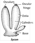
(8) An air (swim) bladder often present with more... 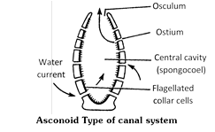 (11) The course taken by the water current way be shown as under –
Ingressing water \[\xrightarrow{\text{Ostia}}\] Spongocoel \[\xrightarrow{\text{Osculum}}\] To outside
(12) The sponges possess an endoskeleton in the form of calcareous spicules.
(13) Excretion and respiration occur by diffusion.
(14) They have greater power of regeneration due to totipotent archaeocytes.
(15) Digestion in sponges is intracellular like protozoans.
(16) All sponges are hermaphrodite, reproduction takes place by asexual or sexual methods.
(17) Gemmules are internal buds containing archaeocytes, mostly found in fresh water sponges, concerned with asexual reproduction.
(18) Development is indirect or direct. The common larval forms are parenchymula (leucosolenia and Clathrina), amphiblastula (Sycon), etc.
Classification of porifera : On the basis of types of endoskeleton, phylum porifera is divisible into three classes
Class 1. Calcarea or Calcispongiae
(1) Skeleton is formed of Calcareous spicules.
(2) Radially symmetrical.
(3) Choanocyte cells are large and conspicuous.
(4) Canal system asconoid (ascon) or syconoid (sycon) type.
(5) These are also known as limy sponges.
Examples : Clathrina, Leucosolenia, Sycon, Grantia, etc.,
(11) The course taken by the water current way be shown as under –
Ingressing water \[\xrightarrow{\text{Ostia}}\] Spongocoel \[\xrightarrow{\text{Osculum}}\] To outside
(12) The sponges possess an endoskeleton in the form of calcareous spicules.
(13) Excretion and respiration occur by diffusion.
(14) They have greater power of regeneration due to totipotent archaeocytes.
(15) Digestion in sponges is intracellular like protozoans.
(16) All sponges are hermaphrodite, reproduction takes place by asexual or sexual methods.
(17) Gemmules are internal buds containing archaeocytes, mostly found in fresh water sponges, concerned with asexual reproduction.
(18) Development is indirect or direct. The common larval forms are parenchymula (leucosolenia and Clathrina), amphiblastula (Sycon), etc.
Classification of porifera : On the basis of types of endoskeleton, phylum porifera is divisible into three classes
Class 1. Calcarea or Calcispongiae
(1) Skeleton is formed of Calcareous spicules.
(2) Radially symmetrical.
(3) Choanocyte cells are large and conspicuous.
(4) Canal system asconoid (ascon) or syconoid (sycon) type.
(5) These are also known as limy sponges.
Examples : Clathrina, Leucosolenia, Sycon, Grantia, etc.,
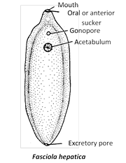 (5) Suctorial pharynx with bifurcated intestine. A large number of caeca or diverticulae arise from each branch of intestine.
(6) Digestion is holozoic. The parasite obtains nourishment from bile, blood, lymph and epithelial cells.
(7) Respiration is anaerobic.
(8) Excretion occurs with the help of flame cells.
(9) Fasciola is a digenetic endoparasite which are its primary host is sheep causing ‘liver rot’ and the secondary or intermediate host is the snail of genus Limnaea and Planorbis.
(10) Fasciola hepatica is a hermaphrodite. In male has a pair of testes and female has an ovary, vitteline gland for yolk formation and mehlis’s gland for lubrication.
(11) Fertilization is internal. Cross fertilization commonly occurs.
(12) Different larval stages of Fasciola hepatica according to development sequence are : miracidium-sporocyst-Redia-Cercaria-Metacercaria.
(13) Stage in the life cycle of Fasciola when it infects intermediate host (snail) is miracidium and primary host is metacercaria.
(14) Miracidium and cercaria larva are free swimming form in water. Redia and sporocyst are formed in snail.
(15) Fasciola exhibits both alternation of generation and alternation of host.
(5) Suctorial pharynx with bifurcated intestine. A large number of caeca or diverticulae arise from each branch of intestine.
(6) Digestion is holozoic. The parasite obtains nourishment from bile, blood, lymph and epithelial cells.
(7) Respiration is anaerobic.
(8) Excretion occurs with the help of flame cells.
(9) Fasciola is a digenetic endoparasite which are its primary host is sheep causing ‘liver rot’ and the secondary or intermediate host is the snail of genus Limnaea and Planorbis.
(10) Fasciola hepatica is a hermaphrodite. In male has a pair of testes and female has an ovary, vitteline gland for yolk formation and mehlis’s gland for lubrication.
(11) Fertilization is internal. Cross fertilization commonly occurs.
(12) Different larval stages of Fasciola hepatica according to development sequence are : miracidium-sporocyst-Redia-Cercaria-Metacercaria.
(13) Stage in the life cycle of Fasciola when it infects intermediate host (snail) is miracidium and primary host is metacercaria.
(14) Miracidium and cercaria larva are free swimming form in water. Redia and sporocyst are formed in snail.
(15) Fasciola exhibits both alternation of generation and alternation of host.
 (3) Sexes are separate with sexual dimorphism. Male is smaller than female with curved tail, two penial setae (copulatory organs) and cloaca. Female is with straight posterior end of the body and posterior transverse anus and separate gonopore situated ventrally 1/3 from the anterior end. In both the excretory pore is situated mid-ventrally, a little behind the mouth. Ventral surface of male bears fifty pairs preanal and five pairs postanal papillae. These sensory papillae are absent in female.
(4) Mouth both in male and female is terminal, triradiate surrounded by three denticulate lips. One median dorsal and two ventrolateral. Dorsal lip bears two sensory double papillae (tangoreceptors). Both sensory papillae and amphids (chemoreceptors) are present on ventrolateral lips.
(5) Body wall consists of outer cuticle, middle epidermis and inner longitudinal muscle layer. Circular layer is absent. Cuticle is thick which is protects the body of the parasite from mechanical injury and also is resistant to action of digestive enzymes of the host. The epidermis is syncytial (coenocytic) with scattered nuclei and without partition walls.
(6) The body cavity of Ascaris is pseudocoel formed by vacuoles originated from persistent embryonic blastocoel.
(7) There is no alimentary canal and digestive gland. The parasite absorbs digested food of the host so their is no need of digestive organs. Absorption occurs through the general body surface. Salivary glands do not occurs in Ascaris.
(8) Respiratory system is absent, respiration is anaerobic.
(9) Excretory system is H-shaped. It is consists of a single excretory cell or renette cell. Excretory products are ammonia and urea.
(10) Sense organs are simple like labial papillae, cervical papillae, anal papillae, amphids and phasmids.
(11) Ascaris is dioecious or unisexual. Testes is single and median, so male Ascaris is monarchic (monodelphic). Only anterior part of testis is functional, so testis (also ovary) is telogonic.
(12) Ascaris sperm is peculiar without flagellum, tail less, asymmetrical and amoeboidal.
(13) Female Ascaris has paired ovaries so female Ascaris is didelphic.
(14) Copulation occurs in the intestine of host. Fertilization in the lower part of uteri. The egg is mammilated, oval, m-shape with three protective covering?outer protein layer, middle chitinous shell and inner membrane made of esterified glycosides.
(15) Embryonic development takes place only outside the body of human host in soil because it requires low temperature, more oxygen and suitable moisture.
(16) Inside the shell the zygote develops into rhabditiform larva or first stage juvenile in 10-14 days.
(17) The larva of first stage is not infective. It rests for a week and completes first more...
(3) Sexes are separate with sexual dimorphism. Male is smaller than female with curved tail, two penial setae (copulatory organs) and cloaca. Female is with straight posterior end of the body and posterior transverse anus and separate gonopore situated ventrally 1/3 from the anterior end. In both the excretory pore is situated mid-ventrally, a little behind the mouth. Ventral surface of male bears fifty pairs preanal and five pairs postanal papillae. These sensory papillae are absent in female.
(4) Mouth both in male and female is terminal, triradiate surrounded by three denticulate lips. One median dorsal and two ventrolateral. Dorsal lip bears two sensory double papillae (tangoreceptors). Both sensory papillae and amphids (chemoreceptors) are present on ventrolateral lips.
(5) Body wall consists of outer cuticle, middle epidermis and inner longitudinal muscle layer. Circular layer is absent. Cuticle is thick which is protects the body of the parasite from mechanical injury and also is resistant to action of digestive enzymes of the host. The epidermis is syncytial (coenocytic) with scattered nuclei and without partition walls.
(6) The body cavity of Ascaris is pseudocoel formed by vacuoles originated from persistent embryonic blastocoel.
(7) There is no alimentary canal and digestive gland. The parasite absorbs digested food of the host so their is no need of digestive organs. Absorption occurs through the general body surface. Salivary glands do not occurs in Ascaris.
(8) Respiratory system is absent, respiration is anaerobic.
(9) Excretory system is H-shaped. It is consists of a single excretory cell or renette cell. Excretory products are ammonia and urea.
(10) Sense organs are simple like labial papillae, cervical papillae, anal papillae, amphids and phasmids.
(11) Ascaris is dioecious or unisexual. Testes is single and median, so male Ascaris is monarchic (monodelphic). Only anterior part of testis is functional, so testis (also ovary) is telogonic.
(12) Ascaris sperm is peculiar without flagellum, tail less, asymmetrical and amoeboidal.
(13) Female Ascaris has paired ovaries so female Ascaris is didelphic.
(14) Copulation occurs in the intestine of host. Fertilization in the lower part of uteri. The egg is mammilated, oval, m-shape with three protective covering?outer protein layer, middle chitinous shell and inner membrane made of esterified glycosides.
(15) Embryonic development takes place only outside the body of human host in soil because it requires low temperature, more oxygen and suitable moisture.
(16) Inside the shell the zygote develops into rhabditiform larva or first stage juvenile in 10-14 days.
(17) The larva of first stage is not infective. It rests for a week and completes first more... 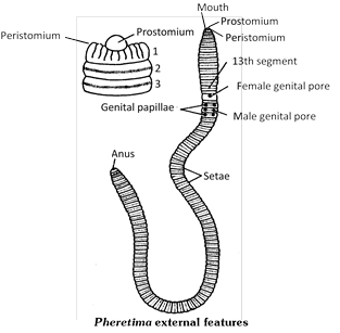 (4) Earthworm is brown or clay-coloured. This is because of the pigment porphyrin. Numerous granules of porphyrin pigment are found scattered in the circular muscle layer of body wall. Porphyrin protects the body from the injurious effects of bright light.
(5) The first segment is peristomium or buccal segment which bears mouth. Anus is located on the last segment.
(6) Three regions in body of earthworm are ? Preclitellar region (1 - 13), Clitellar region (14, 15, 16) and Postclitellar region (17 - last).
(7) Nephridiopores of integumentary nephridia 200-250 per segment found in all segments except the first six. Clitellar segment contains 2000 nephridiopores per segment, so called-forest of nephridia?.
(8) In the body wall 11 pores concerned with reproduction. They are - Spermathecal pores in the intersegmental grooves of 5/6, 6/7, 7/8 and 8/9 (4 pairs). Female genital pore midventral on segment 14th. Male genital pores ventrolaterally (1 pair) on segment 18th.
(9) Male genital papillae are present on segments 17 and 19 (2 pairs).
(10) Body wall is dermomuscular, consists of cuticle, epidermis, muscular layers and coelomic epithelium. Epidermis consists of tall, columnar cells of four types ? Supporting cells (major part), Glandular cells (Goblet and albumin), Basal cells and Sensory cells.
(11) All segments except the first, last and clitellar segment contain setae (perichaetine arrangement). Setae are 'S-shaped, yellowish and chitinous, 80-120 segment. Setae and contraction of muscles help in locomotion.
(12) The body cavity of earthworm is true coelom (schizocoel) as it is formed by the division of mesoderm. The coelom is filled with milky white alkaline coelomic fluid. Coelomic fluid contains different types of carpuscles. These are granulocytes (phagocytes), most numerous mucocytes, circular nucleated cells (leucocytes) and chloragogen cells (yellow cells).
(13) Chloragogen cells are small, star-shaped, yellow cells concerned with storage of reserve food, deamination of proteins, formation of urea and also excretory (analogous to the liver of vertebrates).
(14) The alimentary canal of earthworm is a straight tube, representing a ?tube within tube plan, Location of different part of alimentary canal are -
Buccal chamber : \[1-2\frac{1}{2}\]
Pharynx : \[2\frac{1}{2}-4\]
Oesophagus more...
(4) Earthworm is brown or clay-coloured. This is because of the pigment porphyrin. Numerous granules of porphyrin pigment are found scattered in the circular muscle layer of body wall. Porphyrin protects the body from the injurious effects of bright light.
(5) The first segment is peristomium or buccal segment which bears mouth. Anus is located on the last segment.
(6) Three regions in body of earthworm are ? Preclitellar region (1 - 13), Clitellar region (14, 15, 16) and Postclitellar region (17 - last).
(7) Nephridiopores of integumentary nephridia 200-250 per segment found in all segments except the first six. Clitellar segment contains 2000 nephridiopores per segment, so called-forest of nephridia?.
(8) In the body wall 11 pores concerned with reproduction. They are - Spermathecal pores in the intersegmental grooves of 5/6, 6/7, 7/8 and 8/9 (4 pairs). Female genital pore midventral on segment 14th. Male genital pores ventrolaterally (1 pair) on segment 18th.
(9) Male genital papillae are present on segments 17 and 19 (2 pairs).
(10) Body wall is dermomuscular, consists of cuticle, epidermis, muscular layers and coelomic epithelium. Epidermis consists of tall, columnar cells of four types ? Supporting cells (major part), Glandular cells (Goblet and albumin), Basal cells and Sensory cells.
(11) All segments except the first, last and clitellar segment contain setae (perichaetine arrangement). Setae are 'S-shaped, yellowish and chitinous, 80-120 segment. Setae and contraction of muscles help in locomotion.
(12) The body cavity of earthworm is true coelom (schizocoel) as it is formed by the division of mesoderm. The coelom is filled with milky white alkaline coelomic fluid. Coelomic fluid contains different types of carpuscles. These are granulocytes (phagocytes), most numerous mucocytes, circular nucleated cells (leucocytes) and chloragogen cells (yellow cells).
(13) Chloragogen cells are small, star-shaped, yellow cells concerned with storage of reserve food, deamination of proteins, formation of urea and also excretory (analogous to the liver of vertebrates).
(14) The alimentary canal of earthworm is a straight tube, representing a ?tube within tube plan, Location of different part of alimentary canal are -
Buccal chamber : \[1-2\frac{1}{2}\]
Pharynx : \[2\frac{1}{2}-4\]
Oesophagus more...  (14) Each leg is formed by five segments, viz, coxa, trochanter, femur, tibia and tarsus (tarsus is made by five tarsomeres). Attached to the last tarsomere called pretarsus and it bears, a soft lobe called arolium or pulvilus and a pair of claws is present. They are helpful in moving on smooth surfaces. Plantulae are present on tarsus and act as thermoreceptors.
(15) The most swollen segment in the leg of cockroach is coxa. The longest segment in the leg of cockroach is tibia.
(16) In adult cockroach abdomen is made up of ten segments. But in embryonic stage eleven segments are present. The 11 segment of embryo is represented in adult by podical plates.
(17) In male cockroach, eighth and ninth terga are overlapped by seventh tergum. In female seventh, eighth and ninth sterna are fused to form a brood pouch. Seventh sternum of brood pouch forms a pair of gynavalvular more...
(14) Each leg is formed by five segments, viz, coxa, trochanter, femur, tibia and tarsus (tarsus is made by five tarsomeres). Attached to the last tarsomere called pretarsus and it bears, a soft lobe called arolium or pulvilus and a pair of claws is present. They are helpful in moving on smooth surfaces. Plantulae are present on tarsus and act as thermoreceptors.
(15) The most swollen segment in the leg of cockroach is coxa. The longest segment in the leg of cockroach is tibia.
(16) In adult cockroach abdomen is made up of ten segments. But in embryonic stage eleven segments are present. The 11 segment of embryo is represented in adult by podical plates.
(17) In male cockroach, eighth and ninth terga are overlapped by seventh tergum. In female seventh, eighth and ninth sterna are fused to form a brood pouch. Seventh sternum of brood pouch forms a pair of gynavalvular more... 


You need to login to perform this action.
You will be redirected in
3 sec

