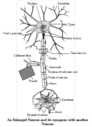
 Types of neurons : Neurons are divided into different categories on different basis.
(1) On the basis of functions : Neurons are divided into three categories :
Sensory (afferent) more...
Types of neurons : Neurons are divided into different categories on different basis.
(1) On the basis of functions : Neurons are divided into three categories :
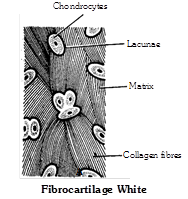
Sensory (afferent) more... 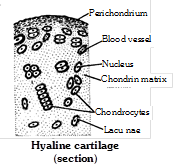 (2) Fibro cartilage (White fibrous cartilage) : In this cartilage, the small amount of matrix of cartilage is packed with large number of bundles of thick white (collagen) fibres. So it is toughest and less flexible. It is found in intervertebral discs and acts as shock absorber. It is also found in pubic symphysis and helps in parturition (child birth). The intervertebral discs remain contracted when the body is active, but relaxed when the body is at rest. That is why, our body becomes a bit taller during sleep and after death.
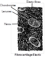
 (3) Elastic cartilage (Yellow elastic cartilage) : In this cartilage, the matrix is packed with yellow or elastic fibres which run in all directions to form a network. Owing to the presence of yellow fibres, it is very flexible. It gives recoiling power to structures. It is found in mammalian pinna, pharyngotympanic tube, epiglottis, some laryngeal and bronchiolar cartilages.
 (4) Calcified cartilage : It is modified hyaline cartilage, It is hard and non elastic due to deposition of calcium salt-hydroxy appetite in matrix. It is found in pubis of old frog, supra-scapula of frog, quadrate cartilage of frog, shark vertebrae, in man ends of long bone, head of humerus and femur. Calcification may also occur as a regular more...
 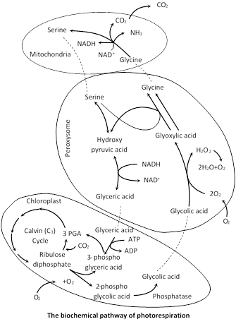 Biochemical mechanism
(1) Ribulose-1, 5-biphosphate \[\xrightarrow{{{O}_{2}}}\] 2 Phoshoglycolic acid + 3 Phoshoglyceric acid
(2) 2 Phosphoglycolic acid \[+{{H}_{2}}O\xrightarrow{Phosphatase}\] Glycolic acid + Phosphoric acid.
(3) Glycolic acid \[+{{O}_{2}}\underset{\text{Oxidase}}{\mathop{\xrightarrow{\text{Glycolic}\,\text{acid}}}}\,\] Glyoxylic acid\[+{{H}_{2}}{{O}_{2}}\] \[2{{H}_{2}}{{O}_{2}}\xrightarrow{\text{Catalase}}2{{H}_{2}}O+{{O}_{2}}\]
(4) Glyoxylic acid + Glutamic acid \[\underset{\text{transaminase}}{\mathop{\xrightarrow{\text{Glutamate}-\text{glyoxylate}}}}\,\]
Glycine \[+\,\,\alpha -\]keto glutaric acid
(5) 2 Glycine \[+{{H}_{2}}O+NA{{D}^{+}}\xrightarrow{{}}\]Serine\[+C{{O}_{2}}+N{{H}_{3}}+NADH\]
(6) Serine + Glyoxylic acid \[\xrightarrow{{}}\] Hydroxypyruvic acid + Glycine Hydroxypyruvic acid \[\xrightarrow{{}}\] Glyceric acid
(7) Glyceric acid + ATP ® 3 phosphoglyceric acid + ADP + phosphate
Importance of photorespiration : Photorespiration is quite different from respiration as no ATP or NADH are produced. Moreover, the process is harmful to plants because as much as half the photosynthetically fixed carbon dioxide (in the form of RuBP) may be lost into the atmosphere through this process.
Any increase in \[{{O}_{2}}\] concentration would favour the uptake of \[{{O}_{2}}\] rather than \[C{{O}_{2}}\] and thus, inhibit photosynthesis for this rubisco functions as RuBP oxygenase. Photorespiration is closely related to \[C{{O}_{2}}\] compensation point and occurs only in those plants which have high \[C{{O}_{2}}\] compensation point such as \[{{C}_{3}}\] plants.
Photorespiration generally occurs in temperate plants. Few photorespiring plants are : Rice, bean, wheat, barley etc. Inhibitors of glycolic acid oxidase such as hydroxy sulphonates inhibit the process of photorespiration. Unlike usual mitochondria respiration neither reduced coenzymes are generated in photorespiration nor the oxidation of glycolate is coupled with the formation of ATP molecules. Photorespiration (\[{{C}_{2}}\] cycle) is enhanced by bright light, high temperature, high oxygen and low \[C{{O}_{2}}\]concentration.
Biochemical mechanism
(1) Ribulose-1, 5-biphosphate \[\xrightarrow{{{O}_{2}}}\] 2 Phoshoglycolic acid + 3 Phoshoglyceric acid
(2) 2 Phosphoglycolic acid \[+{{H}_{2}}O\xrightarrow{Phosphatase}\] Glycolic acid + Phosphoric acid.
(3) Glycolic acid \[+{{O}_{2}}\underset{\text{Oxidase}}{\mathop{\xrightarrow{\text{Glycolic}\,\text{acid}}}}\,\] Glyoxylic acid\[+{{H}_{2}}{{O}_{2}}\] \[2{{H}_{2}}{{O}_{2}}\xrightarrow{\text{Catalase}}2{{H}_{2}}O+{{O}_{2}}\]
(4) Glyoxylic acid + Glutamic acid \[\underset{\text{transaminase}}{\mathop{\xrightarrow{\text{Glutamate}-\text{glyoxylate}}}}\,\]
Glycine \[+\,\,\alpha -\]keto glutaric acid
(5) 2 Glycine \[+{{H}_{2}}O+NA{{D}^{+}}\xrightarrow{{}}\]Serine\[+C{{O}_{2}}+N{{H}_{3}}+NADH\]
(6) Serine + Glyoxylic acid \[\xrightarrow{{}}\] Hydroxypyruvic acid + Glycine Hydroxypyruvic acid \[\xrightarrow{{}}\] Glyceric acid
(7) Glyceric acid + ATP ® 3 phosphoglyceric acid + ADP + phosphate
Importance of photorespiration : Photorespiration is quite different from respiration as no ATP or NADH are produced. Moreover, the process is harmful to plants because as much as half the photosynthetically fixed carbon dioxide (in the form of RuBP) may be lost into the atmosphere through this process.
Any increase in \[{{O}_{2}}\] concentration would favour the uptake of \[{{O}_{2}}\] rather than \[C{{O}_{2}}\] and thus, inhibit photosynthesis for this rubisco functions as RuBP oxygenase. Photorespiration is closely related to \[C{{O}_{2}}\] compensation point and occurs only in those plants which have high \[C{{O}_{2}}\] compensation point such as \[{{C}_{3}}\] plants.
Photorespiration generally occurs in temperate plants. Few photorespiring plants are : Rice, bean, wheat, barley etc. Inhibitors of glycolic acid oxidase such as hydroxy sulphonates inhibit the process of photorespiration. Unlike usual mitochondria respiration neither reduced coenzymes are generated in photorespiration nor the oxidation of glycolate is coupled with the formation of ATP molecules. Photorespiration (\[{{C}_{2}}\] cycle) is enhanced by bright light, high temperature, high oxygen and low \[C{{O}_{2}}\]concentration. 


You need to login to perform this action.
You will be redirected in
3 sec

