Category : 11th Class
The study of blood vessels is called Angiology. The blood vessels are of following types :
(1) Arteries : Thick walled, carrying oxygenated blood (deoxygenated in pulmonary artery) from heart to various parts of body. These blood vessels are grouped as Aorta which branches to form arteries which further divides into thinner branches called arterioles inside the organ. Average diameter of arteriole is \[120\,\,\mu m\] the arterioles further divide into smaller vessels called meta-arterioles \[(70\,\,\mu m)\] which divide into capillaries. At the beginning of capillary, the arterioles posses circular muscles called precapillary sphincter which regulates flow of blood into the capillaries which is called vasomotion. Smooth muscles of arteries innervated by sympathetic fibers, their stimulation control vasoconstriction and vasodilation. Smooth muscles of arteries and arterioles also limit bleeding from wounds by producing vascular spasm during cut. Arteries two types.
(i) Conducting or elastic arteries
(ii) Distributing or muscular arteries.
Elastic or conducting arteries receive blood from heart and do not provide it to any organ rather they provide blood to other atreries and are pressure reservoirs of blood.
Muscleless end of meta-arteriole is called thoroughfare channel or preferential channel.
The largest artery is dorsal / abdominal aorta (systemic aorta).
(2) Capillaries : Smallest blood vessels, discovered by Marcello Malpighi (also layered nucleated squamous epithelial cells called endothelium resting on a basement membrane. Diameter of capillary is about 8m. These are also called as exchange vessels as they are the site of exchange of material between blood and tissue because of least barrier in them. The capillaries can be grouped into two categories :
(i) Arteriolar capillary : Which supplies nutrition, respiratory gases etc. to the body cells.
(ii) Veinular capillaries : Which collect the metabolic wastes from the body cells.
Capillaries possess about 7% of total body blood and are present near almost all cells of body in the intercellular spaces. The tissues which are devoid of intercellular spaces are also devoid of capillary. They are called avascular tissues.
Capillaries are surrounded by cells of connective tissue called pericapillary cells. Some of these cells are contractile and phagocytic in nature and are called Rouget cells or pericytes.
Continuous capillaries are without fenestra/aperture, hence are less permeable. These are present in organs such as lungs, muscles, connective tissues and brain tissues.
Fenestrated capillaries possess apertures/fenestra and are found in those organs where there is maximum need of permeability such as endocrine glands, intestinal villi, cavities of brain, kidney, ciliary body of eye.
Sinusoids are irregularly dilated capillaries found in organs where there is decrease in flow rate such as liver, spleen, bone marrow, parathyroid, pituitary gland. In liver, sinusoids are branches of venules and open into venules while in other organs, they originate from arteriole and unite to form venules.
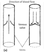
(3) Veins : These are thin walled, carrying deoxygenated blood (oxygenated in pulmonary vein) from tissues to the heart. Venules, smallest branches, unite to form veins which in turn unite to form vena cava. The largest vein is inferior vena cava/post caval. Varicose veins is stout, blood filled painful veins specially of the limbs due to defective watch pocket valves.
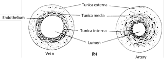
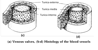
Histology of arteries and veins
(1) Tunica externa or tunica adventitia : Outermost, fibrous, made up of collagen rich connective tissue and less elastin fibres. The collagen fibres give strength to the blood vessels and prevent their overdilation.
(2) Tunica media : Middle, thickest, made up of smooth involuntary muscle fibres and elastin fibres. This layer is very much variable because number of elastin fibres and muscle fibres depend upon the position of blood vessels from the heart.
(3) Tunica interna or tunica intima : Innermost, thinnest, made up of inner, single layer of simple squamous epithelial cells called endothelium resting on a basement membrane and outer layer of elastic (yellow fibrous) connective tissue. The hollow space in the blood vessel is called lumen.
Differences between arteries and veins
|
S.No. |
Characters |
Arteries |
Veins |
|
(1) |
Wall |
Thick, more elastic, non collapsible. |
Thin, less elastic, collapsible. |
|
(2) |
Tunica externa |
Less developed, so less strong. |
More developed, so more strong. |
|
(3) |
Tunica media |
More muscular and has many elastic fibres. |
Less muscular and only a few elastic fibres. |
|
(4) |
Tunica interna |
Endothelial cells more elongated. Elastic membrane more developed. |
Endothelial cells less flat. Elastic membrane less developed. |
|
(5) |
Lumen |
Narrow |
Wider |
|
(6) |
Position |
Deep seated except wrist, neck etc. |
Superficial |
|
(7) |
Valves |
Without valves. |
With valves to prevent back flow. |
|
(8) |
Direction of blood flow |
From heart to body organs |
From body organs to heart |
|
(9) |
Nature of blood |
Oxygenated except pulmonary artery. |
Deoxygenated except pulmonary vein |
|
(10) |
Blood pressure |
More, generally 120/80 mm of Hg. |
Less, generally 0 mm of Hg. |
|
(11) |
Speed of blood |
Fast |
Slow |
|
(12) |
After death |
Becomes empty |
Contain blood |
|
(13) |
Amount of blood |
15% at any given time. |
64% at any given time |
|
(14) |
Colour |
Pink |
Dark red |
|
(15) |
Disintensibility |
Less |
More |
|
(16) |
Average Blood pressure |
More |
Less |
|
(17) |
Elastic-lamina |
Present |
Absent |
Blood pressure : The pressure exerted by the blood on the wall of the blood vessels in which it is present is called blood pressure. It is usually measured in brachial artery by an instrument called sphygmomanometer (invented by Riva-Rocci). Arterial blood pressure is of 2 types :
(1) Systolic blood pressure : It is the pressure exerted by blood on the walls of the blood vessels due to the systole of ventricles and is equal to 120 mm Hg. During ventricular systole, there is expansion in the artery due to the uncoiling of elastic layer. Hence, the pressure is maximum in arteries but gradually decreases in capillaries and veins.
(2) Diastolic blood pressure : It is the pressure exerted on walls of blood vessels when the ventricles are relaxed. During ventricular diastole, the uncoiled elastic layer recoils leading to normalization of artery. Hence, blood pressure drops down to 80 mm Hg. Thus, blood pressure in normal person is systolic/diastolic pressure i.e. 120/80 mm Hg.
(3) Pulse pressure : The difference between systolic and diastolic pressures is called pulse pressure and its normal value is \[12080\,\,mm\text{ }Hg=40\,\,mm\text{ }Hg.\] It provides information about the condition of arteries.
(4) Mean arterial pressure : It is the average pressure of systolic and diastolic pressures. As the blood remains in the systolic phase for shorter period and in the diastolic phase for longer period, the mean pressure of blood lies near the diastolic pressure.
This value varies at different levels of circulation being maximum (100 mm Hg) in the aorta and minimum (0 mm Hg) in the venae cavae under normal conditions.
Pulse : It is the pressure wave of distension and recoiling felt in the radial artery due to the contraction of left ventricle which force about \[70-90\,\,ml\] of blood in each cardiac cycle to aorta. This pressure wave of contraction travels down to the wall to the arteries and is called the pulse.
The pulse is measured in the radial artery in the wrist but can be felt in the temporal artery over the temporal bone or the dorsal pedis artery at the bind of ankle. The pulse normally travels at the rate of 5-8 m/second.
Since each heart beat generates one pulse in the arteries so the pulse rate per minute indicates the rate of heart beat. So the normal pulse rate in a normal adult person is 72/minute.
The normal ratio of systolic pressure to diastolic pressure to pulse pressure is about 3 : 2 : 1.
Factors affecting blood pressure
(1) Age : With the advancing age, BP increases after the age of 60 years, it is calculated as 100 + age of the person.
(2) Cardiac output : BP increases with the increase in cardiac output.
(3) Elasticity of blood vessels : BP is inversely related to the elasticity of the blood vessels.
(4) Total peripheral resistance : Constriction of the blood vessels increases BP whereas dilation of the blood vessels decreases BP.
Hypotension : Low blood pressure with systolic below 110 mm Hg and diastolic below 70 mm Hg. It is caused by low metabolic rate, starvation, anaemia, chronic vasodilation of arterioles, lower pumping activity of heart, loss of blood in haemorrhage, valvular defects, nervous disorders and Addison’s disease. It may cause fainting. Also due to lowering of oxyctosin, Acetylcholine. ANP (Atrial Natriuretic Peptide) Low \[C{{a}^{++}},\]Low BMR.
Hypertension : Persistent high blood pressure with systolic more than 140 mm Hg and diastolic more than 90 mm Hg. It is caused by decrease in extensibility of the artery due to atherosclerosis and arteriosclerosis. Sclerosis means hardening and narrowing of blood vessels which may be due to the deposition of cholesterol or calcium or lipid or any other compound in the wall of the arteries and arterioles.
In atherosclerosis deposition is mainly in tunica interna of the blood vessels which prevents their dilation. The atherosclerosis is, infact, the beginning of thickening and hardening of blood vessels but later, the deposition of cholesterol and other compounds takes places in both tunica media and tunica interna leading to arteriosclerosis.
Hypertension caused by hormones (epinephrine, aldosterone, renin) is called secondary hypertension, other forms of hypertension are known as primary or essential hypertension.


You need to login to perform this action.
You will be redirected in
3 sec

