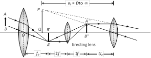
 (1) It consists of three converging lens : objective, eye lens and erecting lens.
(2) It's final image is virtual, erect and smaller.
(3) Magnification : \[{{m}_{D}}=\frac{{{f}_{0}}}{{{f}_{e}}}\left( 1+\frac{{{f}_{e}}}{D} \right)\] and \[{{m}_{\infty }}=\frac{{{f}_{0}}}{{{f}_{e}}}\]
(4) Length : \[{{L}_{D}}={{f}_{0}}+4f+{{u}_{e}}\] and \[{{L}_{\infty }}={{f}_{0}}+4f+{{f}_{e}}\]
(1) It consists of three converging lens : objective, eye lens and erecting lens.
(2) It's final image is virtual, erect and smaller.
(3) Magnification : \[{{m}_{D}}=\frac{{{f}_{0}}}{{{f}_{e}}}\left( 1+\frac{{{f}_{e}}}{D} \right)\] and \[{{m}_{\infty }}=\frac{{{f}_{0}}}{{{f}_{e}}}\]
(4) Length : \[{{L}_{D}}={{f}_{0}}+4f+{{u}_{e}}\] and \[{{L}_{\infty }}={{f}_{0}}+4f+{{f}_{e}}\] 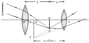 (1) \[{{f}_{\text{objective}}}>{{f}_{\text{eyelens}}}\]and \[{{d}_{\text{objective}}}>{{d}_{\text{eye lens}}}\].
(2) Intermediate image is real, inverted and small.
(3) Final image is virtual, inverted and small.
(4) Magnification : \[{{m}_{D}}=-\frac{{{f}_{0}}}{{{f}_{e}}}\left( 1+\frac{{{f}_{e}}}{D} \right)\] and \[{{m}_{\infty }}=-\frac{{{f}_{o}}}{{{f}_{e}}}\]
(5) Length : \[{{L}_{D}}={{f}_{0}}+{{u}_{e}}\] and \[{{L}_{\infty }}={{f}_{0}}+{{f}_{e}}\]
(1) \[{{f}_{\text{objective}}}>{{f}_{\text{eyelens}}}\]and \[{{d}_{\text{objective}}}>{{d}_{\text{eye lens}}}\].
(2) Intermediate image is real, inverted and small.
(3) Final image is virtual, inverted and small.
(4) Magnification : \[{{m}_{D}}=-\frac{{{f}_{0}}}{{{f}_{e}}}\left( 1+\frac{{{f}_{e}}}{D} \right)\] and \[{{m}_{\infty }}=-\frac{{{f}_{o}}}{{{f}_{e}}}\]
(5) Length : \[{{L}_{D}}={{f}_{0}}+{{u}_{e}}\] and \[{{L}_{\infty }}={{f}_{0}}+{{f}_{e}}\] 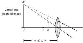 (2) Compound microscope
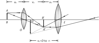 (i) Consist of two converging lenses called objective and eye lens.
(ii) \[{{f}_{\text{eye lens}}}>{{f}_{\text{objective}}}\]and \[{{(\text{diameter)}}_{\text{eye lens}}}>{{(\text{diameter})}_{\text{objective}}}\]
(iii) Intermediate image is real and enlarged.
(iv) Final image is magnified, virtual and inverted.
(v) \[{{u}_{o}}=\]Distance of object from objective (o), \[{{v}_{o}}=\]Distance of image \[({A}'{B}')\] formed by objective from objective, \[{{u}_{e}}=\] Distance of \[{A}'{B}'\] from eye lens, \[{{v}_{e}}=\] Distance of final image from eye lens, \[{{f}_{o}}=\] Focal length of objective, \[{{f}_{e}}=\] Focal length of eye lens.
(vi) Final image is formed at D : Magnification \[{{m}_{D}}=-\frac{{{v}_{o}}}{{{u}_{o}}}\left( 1+\frac{D}{{{f}_{e}}} \right)\] and length of the microscope tube (distance between two lenses) is \[{{L}_{D}}={{v}_{o}}+{{u}_{e}}\].
Generally object is placed very near to the principal focus of the objective hence \[{{u}_{o}}\tilde{=}\,{{f}_{o}}.\] The eye piece is also of small focal length and the image formed by the objective is also very near to the eye piece.
So \[{{v}_{o}}\tilde{=}{{L}_{D}},\] the length of the tube.
Hence, we can write \[{{m}_{D}}=\frac{-L}{{{f}_{o}}}\,\left( 1+\frac{D}{{{f}_{e}}} \right)\]
(vii) Final image is formed at \[\infty \] : Magnification
\[{{m}_{\infty }}=-\frac{{{v}_{0}}}{{{u}_{0}}}.\frac{D}{{{f}_{e}}}\]and length of tube \[{{L}_{\infty }}={{v}_{0}}+{{f}_{e}}\]
In terms of length \[{{m}_{\infty }}=\frac{({{L}_{\infty }}-{{f}_{o}}-{{f}_{e}})D}{{{f}_{o}}{{f}_{e}}}\]
(viii) For large magnification of the compound microscope, both \[{{f}_{o}}\] and \[{{f}_{e}}\] should be small.
(ix) If the length of the tube of microscope increases, then its magnifying power increases.
(x) The magnifying power of the compound microscope may be expressed as \[M={{m}_{o}}\times {{m}_{e}}\]; where \[{{m}_{o}}\] is the magnification of the objective and \[{{m}_{e}}\] is magnifying power of eye piece.
(i) Consist of two converging lenses called objective and eye lens.
(ii) \[{{f}_{\text{eye lens}}}>{{f}_{\text{objective}}}\]and \[{{(\text{diameter)}}_{\text{eye lens}}}>{{(\text{diameter})}_{\text{objective}}}\]
(iii) Intermediate image is real and enlarged.
(iv) Final image is magnified, virtual and inverted.
(v) \[{{u}_{o}}=\]Distance of object from objective (o), \[{{v}_{o}}=\]Distance of image \[({A}'{B}')\] formed by objective from objective, \[{{u}_{e}}=\] Distance of \[{A}'{B}'\] from eye lens, \[{{v}_{e}}=\] Distance of final image from eye lens, \[{{f}_{o}}=\] Focal length of objective, \[{{f}_{e}}=\] Focal length of eye lens.
(vi) Final image is formed at D : Magnification \[{{m}_{D}}=-\frac{{{v}_{o}}}{{{u}_{o}}}\left( 1+\frac{D}{{{f}_{e}}} \right)\] and length of the microscope tube (distance between two lenses) is \[{{L}_{D}}={{v}_{o}}+{{u}_{e}}\].
Generally object is placed very near to the principal focus of the objective hence \[{{u}_{o}}\tilde{=}\,{{f}_{o}}.\] The eye piece is also of small focal length and the image formed by the objective is also very near to the eye piece.
So \[{{v}_{o}}\tilde{=}{{L}_{D}},\] the length of the tube.
Hence, we can write \[{{m}_{D}}=\frac{-L}{{{f}_{o}}}\,\left( 1+\frac{D}{{{f}_{e}}} \right)\]
(vii) Final image is formed at \[\infty \] : Magnification
\[{{m}_{\infty }}=-\frac{{{v}_{0}}}{{{u}_{0}}}.\frac{D}{{{f}_{e}}}\]and length of tube \[{{L}_{\infty }}={{v}_{0}}+{{f}_{e}}\]
In terms of length \[{{m}_{\infty }}=\frac{({{L}_{\infty }}-{{f}_{o}}-{{f}_{e}})D}{{{f}_{o}}{{f}_{e}}}\]
(viii) For large magnification of the compound microscope, both \[{{f}_{o}}\] and \[{{f}_{e}}\] should be small.
(ix) If the length of the tube of microscope increases, then its magnifying power increases.
(x) The magnifying power of the compound microscope may be expressed as \[M={{m}_{o}}\times {{m}_{e}}\]; where \[{{m}_{o}}\] is the magnification of the objective and \[{{m}_{e}}\] is magnifying power of eye piece. 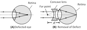 (ii) In this defect focal length or radii of curvature of lens reduced or power of lens increases or distance between eye lens and retina increases.
(iii) This defect can be removed by using a concave lens of suitable focal length.
(iv) If defected far point is at a distance d from eye then Focal length of used lens \[f=-d=-\] (defected far point)
(v) A person can see upto distance \[\to x\], wants to see distance \[\to y(y>x)\] so \[f=\frac{xy}{x-y}\] or power of the lens \[P=\frac{x-y}{xy}\]
(2) Hypermetropia (long sightness) : A long-sighted eye can see distant objects clearly but nearer object are not clearly visible.
(i) Image formed behind the retina and near point moves away
(ii) In this defect focal length or radii of curvature of lens reduced or power of lens increases or distance between eye lens and retina increases.
(iii) This defect can be removed by using a concave lens of suitable focal length.
(iv) If defected far point is at a distance d from eye then Focal length of used lens \[f=-d=-\] (defected far point)
(v) A person can see upto distance \[\to x\], wants to see distance \[\to y(y>x)\] so \[f=\frac{xy}{x-y}\] or power of the lens \[P=\frac{x-y}{xy}\]
(2) Hypermetropia (long sightness) : A long-sighted eye can see distant objects clearly but nearer object are not clearly visible.
(i) Image formed behind the retina and near point moves away
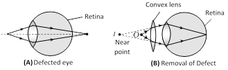 (ii) In this defect focal length or radii of curvature of lens increases or power of lens decreases or distance between eye lens and retina decreases.
(iii) This defect can be removed by using a convex lens.
(iv) If a person cannot see before distance d but wants to see the object placed at distance D from eye so \[f=\frac{dD}{d-D}\] and power of the lens \[P=\frac{d-D}{dD}\]
(3) Presbyopia : In this defect both near and far objects are not clearly visible. It is an old age disease and it is due to the loosing power of accommodation. It can be removed by using bifocal lens.
(4) Astigmatism : In this defect eye cannot see horizontal and vertical lines clearly, simultaneously. It is due to imperfect spherical nature of eye lens. This defect can be removed by using cylindrical lens (Torric lenses).
(ii) In this defect focal length or radii of curvature of lens increases or power of lens decreases or distance between eye lens and retina decreases.
(iii) This defect can be removed by using a convex lens.
(iv) If a person cannot see before distance d but wants to see the object placed at distance D from eye so \[f=\frac{dD}{d-D}\] and power of the lens \[P=\frac{d-D}{dD}\]
(3) Presbyopia : In this defect both near and far objects are not clearly visible. It is an old age disease and it is due to the loosing power of accommodation. It can be removed by using bifocal lens.
(4) Astigmatism : In this defect eye cannot see horizontal and vertical lines clearly, simultaneously. It is due to imperfect spherical nature of eye lens. This defect can be removed by using cylindrical lens (Torric lenses). 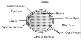 (1) Eye lens : Over all behaves as a convex lens of \[\mu =1.437\]
(2) Retina : Real and inverted image of an object, obtained at retina, brain sense it erect.
(3) Yellow spot : It is the most sensitive part, the image formed at yellow spot is brightest.
(4) Blind spot : Optic nerves goes to brain through blind spot. It is not sensitive for light.
(5) Ciliary muscles : Eye lens is fixed between these muscles. It's both radius of curvature can be changed by applying pressure on it through ciliary muscles.
(6) Power of accomodation : The ability of eye to see near objects as well as far objects is called power of accomodation.
(7) Range of vision : For healthy eye it is 25 cm (near point) to \[\infty \] (far point).
A normal eye can see the objects clearly, only if they are at a distance greater than 25 cm. This distance is called Least distance of distinct vision and is represented by D.
(8) Persistence of vision : Is 1/10 sec. i.e. if time interval between two consecutive light pulses is lesser than 0.1 sec., eye cannot distinguish them separately.
(9) Binocular vision : The seeing with two eyes is called binocular vision.
(10) Resolving limit : The minimum angular separation between two objects, so that they are just resolved is called resolving limit. For eye it is \[{{1}^{'}}={{\left( \frac{1}{60} \right)}^{o}}\].
(1) Eye lens : Over all behaves as a convex lens of \[\mu =1.437\]
(2) Retina : Real and inverted image of an object, obtained at retina, brain sense it erect.
(3) Yellow spot : It is the most sensitive part, the image formed at yellow spot is brightest.
(4) Blind spot : Optic nerves goes to brain through blind spot. It is not sensitive for light.
(5) Ciliary muscles : Eye lens is fixed between these muscles. It's both radius of curvature can be changed by applying pressure on it through ciliary muscles.
(6) Power of accomodation : The ability of eye to see near objects as well as far objects is called power of accomodation.
(7) Range of vision : For healthy eye it is 25 cm (near point) to \[\infty \] (far point).
A normal eye can see the objects clearly, only if they are at a distance greater than 25 cm. This distance is called Least distance of distinct vision and is represented by D.
(8) Persistence of vision : Is 1/10 sec. i.e. if time interval between two consecutive light pulses is lesser than 0.1 sec., eye cannot distinguish them separately.
(9) Binocular vision : The seeing with two eyes is called binocular vision.
(10) Resolving limit : The minimum angular separation between two objects, so that they are just resolved is called resolving limit. For eye it is \[{{1}^{'}}={{\left( \frac{1}{60} \right)}^{o}}\].  Line emission spectrum
(i) It consist of distinct bright lines.
(ii) It is produced by an excited source in atomic state.
(iii) e.g. Spectrum of excited helium, mercury vapours, sodium vapours or atomic hydrogen.
Line emission spectrum
(i) It consist of distinct bright lines.
(ii) It is produced by an excited source in atomic state.
(iii) e.g. Spectrum of excited helium, mercury vapours, sodium vapours or atomic hydrogen.
 Band emission spectrum
(i) It consist of district bright bands.
(ii) It is produced by an excited source in molecular state.
(iii) e.g. Spectra of molecular \[{{H}_{2}},\] CO, \[N{{H}_{3}}\] etc.
Band emission spectrum
(i) It consist of district bright bands.
(ii) It is produced by an excited source in molecular state.
(iii) e.g. Spectra of molecular \[{{H}_{2}},\] CO, \[N{{H}_{3}}\] etc.
 (2) Absorption spectrum : When white light passes through a semi-transparent solid, or liquid or gas, it's spectrum contains certain dark lines or bands, such spectrum is called absorption spectrum (of the substance through which light is passed).
(i) Substances in atomic state produces line absorption spectra. Polyatomic substances such as \[{{H}_{2}},\]\[C{{O}_{2}}\] and \[KMn{{O}_{4}}\]produces band absorption spectrum.
(ii) Absorption spectra of sodium vapour have two (yellow lines) wavelengths \[{{D}_{1}}(5890\,{\AA})\] and \[{{D}_{2}}(5896\,{\AA})\]
(3) Fraunhoffer's lines : The central part (photosphere) of the sun is very hot and emits all possible wavelengths of the visible light. However, the outer part (chromosphere) consists of vapours of different elements. When the light emitted from the photosphere passes through the chromosphere, certain wavelengths are absorbed. Hence, in the spectrum of sunlight a large number of dark lines are seen called Fraunhoffer lines.
(2) Absorption spectrum : When white light passes through a semi-transparent solid, or liquid or gas, it's spectrum contains certain dark lines or bands, such spectrum is called absorption spectrum (of the substance through which light is passed).
(i) Substances in atomic state produces line absorption spectra. Polyatomic substances such as \[{{H}_{2}},\]\[C{{O}_{2}}\] and \[KMn{{O}_{4}}\]produces band absorption spectrum.
(ii) Absorption spectra of sodium vapour have two (yellow lines) wavelengths \[{{D}_{1}}(5890\,{\AA})\] and \[{{D}_{2}}(5896\,{\AA})\]
(3) Fraunhoffer's lines : The central part (photosphere) of the sun is very hot and emits all possible wavelengths of the visible light. However, the outer part (chromosphere) consists of vapours of different elements. When the light emitted from the photosphere passes through the chromosphere, certain wavelengths are absorbed. Hence, in the spectrum of sunlight a large number of dark lines are seen called Fraunhoffer lines.
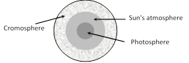 (i) The prominent lines in the yellow part of the visible spectrum were labelled as D-lines, those in blue part as F-lines and in red part as C-line.
(ii) From the study of Fraunhoffer's lines the presence of various elements in the sun's atmosphere can be identified e.g. abundance of hydrogen and helium.
(iii) In the event of a solar eclipse, dark lines become bright. This is because of the reason that the presence of an opaque obstacle in between sun and earth cuts the light off from the central region (photo-sphere), while light from corner portion (cromosphere) is still being received. The bright lines appear exactly at the places where dark lines were present.
(4) Spectrometer : A spectrometer is used for obtaining pure spectrum of a source in laboratory and calculation of \[\mu \] of material of prism and \[\mu \] of a transparent liquid.
It consists of three parts : Collimator which provides a parallel beam of light; Prism Table for holding the prism and Telescope for observing the more...
(i) The prominent lines in the yellow part of the visible spectrum were labelled as D-lines, those in blue part as F-lines and in red part as C-line.
(ii) From the study of Fraunhoffer's lines the presence of various elements in the sun's atmosphere can be identified e.g. abundance of hydrogen and helium.
(iii) In the event of a solar eclipse, dark lines become bright. This is because of the reason that the presence of an opaque obstacle in between sun and earth cuts the light off from the central region (photo-sphere), while light from corner portion (cromosphere) is still being received. The bright lines appear exactly at the places where dark lines were present.
(4) Spectrometer : A spectrometer is used for obtaining pure spectrum of a source in laboratory and calculation of \[\mu \] of material of prism and \[\mu \] of a transparent liquid.
It consists of three parts : Collimator which provides a parallel beam of light; Prism Table for holding the prism and Telescope for observing the more... 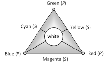 (i) Complementary colours : Green and Magenta, Blue and Yellow, Red and Cyan.
(ii) Combination : Green + Red + Blue = White, Blue + Yellow = White, Red + Cyan = White, Green + Magenta = White
(6) Colour triangle for pigment and dyes : Red, Yellow and Blue are the primary colours.
(i) Complementary colours : Green and Magenta, Blue and Yellow, Red and Cyan.
(ii) Combination : Green + Red + Blue = White, Blue + Yellow = White, Red + Cyan = White, Green + Magenta = White
(6) Colour triangle for pigment and dyes : Red, Yellow and Blue are the primary colours.
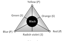 (i) Complementary colours : Yellow and Mauve, Red and Green, Blue and Orange.
(ii) Combination : Yellow + Red + Blue = Black, Blue + Orange = Black, Red + Green = Black, Yellow + Mauve = Black
(i) Complementary colours : Yellow and Mauve, Red and Green, Blue and Orange.
(ii) Combination : Yellow + Red + Blue = Black, Blue + Orange = Black, Red + Green = Black, Yellow + Mauve = Black 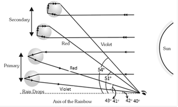 (1) Primary rainbow : (i) Two refraction and one TIR. (ii) Innermost arc is violet and outermost is red. (iii) Subtends an angle of \[{{42}^{o}}\]at the eye of the observer. (iv) More bright
(2) Secondary rainbow : (i) Two refraction and two TIR. (ii) Innermost arc is red and outermost is violet. (iii) It subtends an angle of \[{{52.5}^{o}}\]at the eye. (iv) Comparatively less bright.
(1) Primary rainbow : (i) Two refraction and one TIR. (ii) Innermost arc is violet and outermost is red. (iii) Subtends an angle of \[{{42}^{o}}\]at the eye of the observer. (iv) More bright
(2) Secondary rainbow : (i) Two refraction and two TIR. (ii) Innermost arc is red and outermost is violet. (iii) It subtends an angle of \[{{52.5}^{o}}\]at the eye. (iv) Comparatively less bright. 


You need to login to perform this action.
You will be redirected in
3 sec

