Category : UPSC
The Nervous System
Divisions of the Nervous System
Nervous System in Various Organisms
Coelenterates, cnidarians, and echinoderms have their neurons organized into a nerve net. These creatures have radial symmetry and lack a head. Although lacking a brain or either nervous system (CNS or PNS) nerve nets are capable of some complex behavior. Bilaterally symmetrical animals have a body plant that includes a defined head and a tail region. Development of bilateral symmetry is associated with cephalization of sensory organs at the front end of the organism. Flatworms have neurons associated into clusters known as ganglia, which in turn form a small brain. Vertebrates have a spinal cord in addition to a more developed brain. Chordates have a dorsal rather than ventral nervous system. Several evolutionary trends occur in chordates: spinal cord, continuation of cephalization in the form of larger and more complex brains, and development of a more elaborate nervous system.
The Neuron
Nervous tissue is composed of two main cell types: neurons and glial cells. Neurons transmit nerve messages. Glial cells are in direct contact with neurons and often surround them.
The neuron is the functional unit of the nervous system. Humans have about 100 billion neurons in their brain alone! While variable in size and shape,
a. Parts of Neuron:
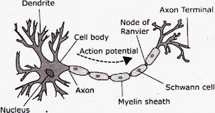
All neurons have three parts.
Dendrites receive information from another cell and transmit the message to the cell body.
The cell body contains the nucleus, mitochondria and other organelles typical of eukaryotic cells.
The axon conducts messages away from the cell body.
b. Types of Neuron:
Three types of neutrons occur, Sensory neurons typically have a long dendrite and short axon, and carry messages from sensory receptors to the central nervous system.
Motor neurons have a long axon and short dendrites and transmit messages from the central nervous system to the muscles (or to glands).
Interneurons are found only in the central nervous system where they connect neuron to neuron. Some axons are wrapped in a myelin sheath formed from the plasma membranes of specialized glial cells known as Schwann cells. Schwann cells serve as supportive, nutritive, and service facilities for neurons. The gap between Schwann cells in known as the node of Ranvier, and serves as points along the neuron for generating a signal. Signals jumping from node to node travel hundreds of times faster than signals traveling along the surface of the axon. This allows our brain to communicate with our toes in a few thousandths of a second.
The Nerve Message
The plasma membrane of neurons, like all other cells, has an unequal distribution of ions and electrical charges between the two sides of the membrane. The outside of the membrane has a positive charge, inside has a negative charge.
Resting potential results from differences between sodium and potassium positively charged ions and negatively charged ions in the cytoplasm.
Sodium ions are more concentrated outside the membrane, while potassium ions are more concentrated inside the membrane. This imbalance is maintained by the active transport of ions to reset the membrane known as the sodium potassium pump.
The sodium-potassium pump maintains this unequal concentration by actively transporting ions against their concentration gradients. The action potential begins at one spot on the membrane, but spreads to adjacent areas of the membrane, propagating the message along the length of the cell membrane. After passage of the action potential, there is a brief period, the refractory period, during which the membrane cannot be stimulated. This prevents the message from being transmitted backward along the membrane.
STEPS IN AN ACTION POTENTIAL
Synapses
The junction between a nerve cell and another cell is called a synapse. Messages travel within the neuron as an electrical action potential. The space between two cells is known as the synaptic cleft. To cross the synaptic cleft requires the actions of neurotransmitters. Neurotransmitters are stored in small synaptic vesicles clustered at the tip of the axon. Neurotransmitters tend to be small molecules, some are even hormones. The neurotransmitters cross the cleft, binding to receptor molecules on the next cell, prompting transmission of the message along that cell’s membrane. Diseases that effect the function of signal transmission can have serious consequences. Parkinson’s disease has a deficiency of the neurotransmitter dopamine. Progressive death of brain cells increases this deficit, causing tremors, rigidity and unstable posture.
Endocrine System
You will know it in detail Chapter five.
Nervous System: The Telegraphic System of Communication and Co-Ordination


You need to login to perform this action.
You will be redirected in
3 sec

