The methods of reproduction which do not involve meiosis and fertilization are known as apomixis or asexual reproduction. Only mitotic divisions are involved in these methods, resulting into the formation of offsprings which are genetically similar to the parent plant.
Asexual reproduction is of following two types :
(1) Agamospermy : Agamospermy is a kind of plant apomixis in which the embryos and seeds are formed by asexual reproductive methods without involving meiotic gametogenesis and sexual fusion of gametes. It occurs widely in ferns and the flowering plants. There are three different types of agamospermy :
(i) Adventive embryony : Formation of embryo directly from the diploid sporophytic cells (nucellus or integument) of ovule is called adventive embryony. Such embryos are formed without involving meiosis and sexual fusion, e.g., Citrus, Opuntia, etc. In Citrus, a seed may possess upto 40 embryos (one normal and rest adventive).
(ii) Diplospory : In this case, the archesporium differentiates but megaspore mother cell directly gives rise to an unreduced (i.e., without meiosis) embryo sac. It may produce two types of embryos:
(a) Diploid parthenogenesis : Embryo develops from unfertilized diploid egg.
(b) Diploid apogamy : Embryo develops from any diploid cell of embryo sac except egg.
(iii) Apospory : It is the formation of complete embryo sac from the sporophytic cell without meiosis so that the gametophyte remains diploid. Apospory may be of two types :
(a) Somatic apospory : Embryo sac is formed from somatic cell.
(b) Generative apospory : Embryo sac is formed from archesporium without meiosis.
(2) Vegetative propagation : Regeneration or Formation of a new individual from any vegetative part of the body is called vegetative reproduction or vegetative propagation. The lower plants reproduce vegetatively through budding, fission, fragmentation, gemmae, resting buds, spores, etc. It is very common mode of reproduction and it may be natural vegetative propagation or artificial vegetative propagation.
(i) Natural methods of vegetative propagation : In natural vegetative propagation, a portion gets deattached from the body of mother plant and it grows into a new individual plant under suitable conditions. Different plant parts are variously modified for vegetative propagation. Some of these are given below.
(a) Vegetative propagation by stems : The modified stems like bulbs, runners, rhizomes, corms, tubers, offsets, etc., help the plant to multiply under favourable conditions.
(b) Vegetative propagation by roots : The roots of some woody plants produce shoots which grow into new plants; e.g., Murraya, Lebbeck tree (Albizzia), Sisham (Dalbergia sisso), etc. Modified tuberous roots of Sweet potato, Asparagus, Dahlia, Tapioca, Tinospora, etc. develop buds and each of which form a new plant.
(c) Vegetative propagation by leaves : The leaves generally do not help in vegetative propagation. However, in Bryophyllum pinnatum and B. daigremontianum, develop along the leaf margins which on deattachment produce independent plants. In elephant ear plant (Begonia) also, leaf buds are produced from petiole and veins throughout the surface of the leaf.
(d) Vegetative propagation by reproductive parts : Flowers are primarily associated with sexual reproduction. more...
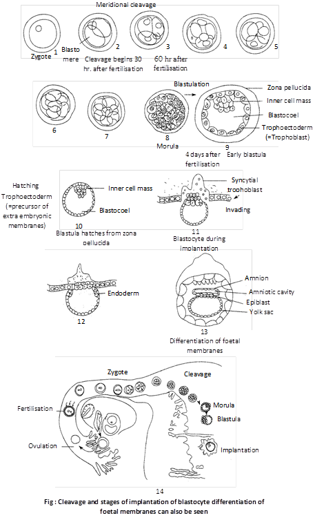
Definition : The term cleavage refers to a series of rapid mitotic division of the zygote following fertilization, forming a many celled blastula. The cleavage follows fertilization and ends with the formation of a characteristic development stage called blastula.
Cleavage versus typical mitosis : The cleavage division are no doubt mitotic as they produce diploid cells, they differ from typical mitosis in a couple of significant points.
Different between cleavage and mitosis
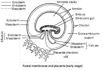
An aquatic embryo is surrounded by water, which protects the embryo, keep it moist, removes wastes and permits gas exchange. In land vertebrate (reptiles, birds and mammals), these functions are taken over by the extra embryonic membranes. These membranes are formed outside the embryo from the trophoblastic only in amniotes and perform specific function. Some of these membranes take part in the formation of placenta in mammals.
(1) Yolk sac : It is formed below the embryo. It contains fluid, not yolk. The yolk sac is a vestigeal organ inherited from the oviparous reptilian ancestors. Yolk sac encloses by outer mesoderm and inner endodermal layer.
Function : In human beings, it is vestigial. In human embryo it act as the site of blood cell formation until about the 6th week, when the liver takes over this role.
(2) Amnion : It is formed above the embryo. It consist of outer mesoderm and inner ectoderm. The amnion and the fluid filled amniotic cavity it encloses, enlarge and nearly surround the embryo. Amniotic fluid secreted by both embryo and amnion.
Functions
(i) The amniotic fluid cushions the embryo.
(ii) It protects the embryo from jerk, injury and shocks.
(iii) It prevents desiccation of the embryo.
(3) Allantois : It is a fold of splanchnopleur developed from the hind gut of the embryo. It consist of outer mesoderm and inner endoderm.
Functions
(i) The cavity of the allantois serves as a urinary bladder. It stores the protein breakdown product in the form of water-insoluble crystals of uric acid and inside the egg upto the time of hatching.
(ii) The vascular “chorioallantoic membrane” lies in a close proximity to the inner surface of the porous shell. It acts as an extraembryonic lung by supplying the embryo with oxygen.
(4) Chorion : It is outermost fold of somatopleur (outer ectoderm and somatic mesoderm) and surrounds the embryo. In reptiles, birds and prototherians, allantochorion act as extra embryonic lungs help in exchange of gases. But in primates including human beings, only chorion forms the placenta (chorionic placenta).
Function : It protects the embryo and forms placenta for metabolic exchange between the foetus and the mother.
S. No.
Name of membrane
Characteristics and functions
Remarks
(1)
Yolk sac
(1) Formed by inner endoderm and outer mesoderm (= splanchnopleura)
(2) Digestive function (= extra embryonic duct)
(3) Absorbs dissolved yolk and supplicate it to developing embryo.
Vestigeal in humans.
Well developed in reptiles, bird and prototherians.
(2)
Amnion
(1) Formed by inner ectoderm and outer mesoderm (somatopleur) above the embryo.
(2) Between the embryo and amnion there is a cavity called more...
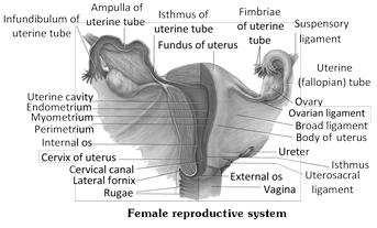
The female reproductive system consists of a pair of ovaries, a pair of fallopian tubes, uterus, vagina, external genitalia or vulva and breasts.
Reproductive organ
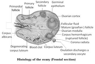
(1) Ovaries : Ovaries are the primary sex organs of female.
The ovaries are almond shaped bodies, about 3 cm long, 1.5 cm wide and 1 cm thick. The ovaries, like the testes, have both an exocrine function (production of ova) and an endocrine role (secretion of female sex hormones : estrogen and progesterone). After menopause, the ovaries become small and lose follicles.
Each ovary is located close to the lateral walls of the pelvic cavity, being suspended from the dorsal body wall just behind the kidney, by a section of peritonium, the mesovarium.
Each ovary is a compact or solid organ, consisting of an outer cortex and inner medulla. The stroma of the cortical region is composed of spindle shaped fibroblasts. A poorly delineated dense connective tissue layer, the tunica albuginea, covers the cortex. It imparts the whitish colour to the ovary. Located outside the tunica albuginea, the germinal epithelium, formed of simple squamous or cuboidal epethelial cells, covers the surface of the ovary.
(2) Fallopian tubes / Uterine tubes / Oviducts : Each ovary is located in front of a funnel shaped opening of the uterus, the oviduct. The oviduct is a muscular tube, measuring about 12 cm in length. Its lumen is lined by ciliated epithelium.
Oviducts develop from the mullarian duct of the embryo. It conveys the egg from the ovary to the uterus, and provides the appropriate environment for its fertilization. It is supported by a double fold of peritoneum called mesosalpinx. The wall of oviduct is made of three layers :
(i) Serosa : It is the outermost layer of visceral-peritoneum.
(ii) Muscle-layer : The middle layer of the oviduct is made up of unstriped-muscle.
(iii) Mucous membrane : It is the innermost layer. This layer is made up of ciliated columnar epithelium and the connective tissue.
The oviduct shows 4 regions :
(a) Infundibulum : It is the broad, funnel-shaped proximal part. Its margin bears motile, finger-like processes called fimbriae. It opens into the body cavity by an aperture called ostium. The latter lies close to the ovary to receive the egg released from the ovary. The fimbriae bear cilia which beat toward the ostium to direct the egg into the infundibulum.
(b) Ampulla : It is the long, wide, thin-walled, tortuous major part of the fallopian tube next to the infundibulum. Ampulla is site for fertilization.
(c) Isthmus : It is the very short, narrow, thick-walled, straight part that follows the ampulla.
(d) Uterine part : It is also narrow and passes through the uterine wall, and communicates with the uterine cavity.
(3) Uterus : It is pyriform, hollow muscular thick-walled but distensible median structure more...
Definition : Fusion of a haploid male gamete (spermatozoan) and a haploid female gamete (ovum) to form a diploid cell, the zygote, is called fertilization or syngamy.
Site of fertilization : Fertilization in human female is internal as in other mammals. It takes place usually in the ampulla of the fallopian tube.
Steps of fertilization
(1) Approach of sperm to ovum : Male discharge semen (3.5 ml) high up in the female’s vagina, close to the cervix during coitus. This is called ejaculation or insemination. This ejaculation contains as many as 400 million sperms but only about 100 sperms reach the fallopian tube because many sperms are either killed by the acidity of female genital tract or engulfed by the phagocytes of the vaginal epithelium. The sperm swim in the seminal fluid at the rate of 1-4 mm per minute by the aspiratory action of the uterus and peristaltic movement of the fallopian tube.
Capacitation is the phenomenon of physiological maturation of sperms by breaking of acrosome membrane inside the female genital tract. It takes about 5-6 hours.
(2) Penetration of sperm : The ovum secretes a chemical substance called fertilizin, which has a number of spermophillic sites on its surface where the sperm of species specific type can be bound by their antifertilizin site. This fertilizin-antifertilizin interaction, causing agglutination (sticking together) of egg and sperm.
Penetration of sperm is a chemical mechanism. In this acrosome of sperm undergoes acrosomal reaction and releases certain sperm lysins which dissolve the egg envelopes locally and make the path for the penetration of sperm. Sperm lysins are acidic proteins. These sperm lysins contain a lysing enzyme hyaluronidase which dissolves the hyaluronic acid polymers in the intercellular spaces which holds the granulosa cells of corona radiata together; corona penetrating enzyme (that dissolves the corona radiata) and acrosin (which dissolves the zona pellucida).
(3) Cortical reaction : Immediately after the entry of a sperm into the egg, the later shows a cortical reaction to check the entry of more sperms. In this reaction, the cortical granules present beneath the egg’s plasma membrane release chemical substance between the ooplasm and the plasma membrane (vitelline membrane).
Sperm penetration into ovum also induces following metabolic activities :
(i) The egg surface produces fertilization cone.
(ii) The vitelline membrane is lifted and is converted into fertilization membrane.
(iii) The cytoplasm exhibits movements.
(iv) The permeability of plasma membrane increases.
(v) The rate of protein synthesis increases.
(vi) Mitosis is initiated.
(4) Fusion of gametic nuclei : Entrance of spermatozoan serves to acts as stimulus which causes the second maturation division. As the head and middle piece of the sperm advance into the egg, those parts rotate through an angle of 180° so that the mitochondria and proximal centriole of the associated middle piece assume the leading position. The centriole brought in by the spermatozoan subdivides into two and as achromatic spindle is established more...
The process of the formation of haploid gametes from the undifferentiated, diploid germ cells in the gonads for sexual reproduction is called gametogenesis.
The process of Gametogenesis is stimulated by the FSH or Follicle Stimulating Hormone and for this process Vitamin "A" and "E" are also necessary.
As a result of this process, male gamete sperm and female gamete egg is formed.
Types of gametogenesis
(1) Spermatogenesis
The process of formation of sperms in seminiferous tubules of the testis of the male animal is termed as spermatogenesis.
In mammals, testis have several coiled tubules in it called the seminiferous tubules. Sperms are formed in these tubules. The inner wall of seminiferous tubules is made up of germinal epithelium whose cells are cuboidal.
The endodermal cells of yolk sac migrate in testes and become primordial germ cells. Due to the division of these cells sperms are formed.
Some large cells are also found in this germinal epithelium. These are called the "Sertoli cells or Sustentacular cells". These cells provide nutrition to the maturing sperms in the form of Glycogen. For getting nutrition, the head of the sperms are submerged in the cytoplasm of sertoli cells.
Sertoli cells mainly provide nutrition and conserve the various stages of spermatogenesis. Spermatogenesis is a continuous process. To make it easier for study, it has been divided into the following steps -
(i) Formation of spermatid.
(ii) Spermiogenesis or Spermateleosis.
(i) Formation of spermatids : This process begins as the animal attains sexual maturity. The endodermal cells of the yolk sac which participate in this process are termed as the primordial germ cells. The process of formation of spermatids from primordial germ cells are termed as spermatocytosis. It has 3 sub-stages -
(a) Multiplication phase : During this process the primordial germ cells repeatedly undergo mitosis division, and as a result of these divisions spermatogonia are formed. Spermatogonia are diploid.
(b) Growth phase : Some spermatogonia either due to growth or due to food storage become 2 or 3 times of their original size, and are now known as primary spermatocytes. The remaining spermatogonia remain in the seminiferous tubules in the form of reserved stock. The primary - spermatocytes formed during the growth phase are diploid. Growth phase is the longest.
(c) Maturation phase : Primary - spermatocytes undergo Meiosis-I and as a result 2 haploid secondary spermatocytes are formed. This division is termed as First Maturation Division or Reductional division. Secondary spermatocytes undergo Meiosis II or equational division, and as result, 2 spermatids are formed from each secondary spermatocyte. Thus, from 1 diploid primary spermatocytes 2 secondary spermatocytes are formed on meiosis I and from 2 haploid secondary spermatocytes 4 spermatids are formed on meiosis-II. Metamorphosis of spermatids into sperms in known as Spermiogenesis or Spermatoliosis.
(ii) Spermatoliosis : The process of transformation of a round non-motile and haploid spermatid obtained from spermatocytosis into thread-like, motile and haploid sperm more...
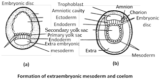
Definition : Gastrulation is a dynamic process involving critical changes in the embryo such as differentiation of cells, establishment of the three primary germ layers and transformation of the single walled blastula into a double walled gastrula.Types of gastrular movement or morphogenetic movement : The movements of cells during gastrulation is called formative or morphogenetic movements. Following types of gastrular movements are found in different animals(1) Epiboly : It involves the morphogenetic movement of prospective ectodermal (micromeres) blastomeres antero-posteriorly to envelop the presumptive endodermal and mesodermal blastomeres. It is found in telolecithal egg of frog.(2) Emboly : It involves inward movement of prospective endodermal and chorda-mesodermal blastomeres from the surface of blastula. Emboly includes following methods :(i) Invagination : It involves insinking of endodermal cells in the blastocoel to form archenteron. It is found in amphioxus.(ii) Involution : It involves the rolling in of the chorda-mesodermal blastomeres inside the ectodermal cells over the lips of blastopore. It is also found in the gastrulation of frog.(iii) Ingression or polyinvagination : In this, individual blastomeres migrate into the blastocoel either from only vegetal pole (called unipolar ingression e.g., Obelia;) or from all sides (called multipolar ingression e.g., Hydra) to form a solid gastrula called stereogastrula.(iv) Delamination : It involves splitting off the blastoderm into two layers by the appearance of grooves resulting the formation of hypoblast. It is found in birds.Formation of layers by gastrulation : Gastrulation includes the formation of following structures(1) Formation of endoderm : The blastodermic vesicle enlarges and cells present on the lower surface of the embryonal knob detach by delamination from the embryonal knob. The part of endoderm located under the embryonal knob is called embryonic endoderm which later forms embryonic gut, while the remaining part of endoderm along with trophoblast forms the yolk sac.(b) Formation of embryonic disc and mesoderm : Meanwhile, the blastocyst continues to grow due to absorption of more and more uterine milk. The embryonal knob stretches and cells of Rauber start breaking off and dispersing. So the cells of embryonal knob from a regular layer called embryonic disc which becomes continuous with the trophoblast. Embryonic disc is differentiated into cephalic, embryonic and caudal regions. Formation of embryonic mesoderm starts at the caudal region of the embryonic disc where cells undergo rapid proliferation and form a localized thickening of the embryonic disc and form the mesodermal layer between ectoderm and endoderm.(3) Formation of ectoderm : The remaining cells of blastodisc become columnar and form ectoderm.Fate of germ layers : Each of more...
(i) Definition: The process of attachment of the blastocyst on the endometrium of the uterus is called implantation.
(ii) Period: Though the implantation may occur at any period between 6th and 10th day after the fertilization but generally it occurs on seventh day after fertilization.
(iii) Mechanism: First of all, the blastocyst is held closely against the uterine endometrial epithelium. The uterine capillaries and uterine wall in the immediate vicinity of the embryo become more permeable and a local stromal edema is developed. Soon the endometrium around the embryo shows the first sign of a decidual cell reaction (DCR) which involves:
(a) The epithelium becomes disrupted and the loosely packed fibroblast-like cells of the stoma are transformed into large rounded glycogen-filled cells.
(b) The area of contact becomes more vascular.
(c) The decidual cells form an “implantation chamber” around the embryo before the formation of a functional placenta.
(d) The tropho blast is developed from the superficial layer of the morula stage. Later, the trophoblast is lined by mesoderm to form the chorion which contributes to the placenta formation.
(e) Trophoblast of the chorion penetrates the uterine epithelium by both cytolytic and mechanical activity. The phagocytic activity of the trophoblastic cells through the decidual cells continues till it establishes intimate connection with the uterine blood vessels. The process of implantation is aided by proteolytic enzymes produced by the trophoblast. After implantation, endometrium undergoes many changes and forms decidua. It is differentiates into three parts such as : Decidua basalis present between the embryo and uterine myometrium, Decidua capsularis lies between the embryo and lumen of the uterus and Decidua parietalis is formed by the remaining part of decidua. The pattern of implantation of the blastocyst varies in different species, which are as follows
(1) Interstitial implantation: The blastocyst get burried into the endometrium e.g. human female, hedgehog, guinea pig, some bats and ape.
(2) Central implantation: The blastocyst remain the uterine cavity e.g. rabbit, cow, dog and monkey.
(3) Eccentric implantation: The blastocyst comes to lie in a uterine recess e.g. rats, mice.
(iv) Hormonal control of implantation
(a) Role of estrogens: These are a group of steroid hormones mainly secreted by follicular epithelial cells of Graafian follicle though these are also produced by adrenal cortex and placenta. These include b-estradiol, esterone, estriol etc. Out of which most important estrogen is b-estradiol. Secretion of estrogens is stimulated by FSH of anterior lobe of pituitary glands. These stimulate the uterine endometrial epithelium to enlarge, become more vascular and more glandular. The uterine glands become tortuous and cork-screw shaped. So the endometrium prepares itself for implantation. This stimulation by the estrogens on the uterus generally occurs on the 4th day of pregnancy.
(b) Progesterone: It is also a steriod hormone secreted by yellow-coloured endocrine gland, called corpus luteum, formed from empty Graafian folicle during the pregnancy. Small amount of progesterone is also secreted by adrenal cortex and placenta. Secretion of progesterone is more...
 Function : It protects the embryo and forms placenta for metabolic exchange between the foetus and the mother.


 (2) Fallopian tubes / Uterine tubes / Oviducts : Each ovary is located in front of a funnel shaped opening of the uterus, the oviduct. The oviduct is a muscular tube, measuring about 12 cm in length. Its lumen is lined by ciliated epithelium.
Oviducts develop from the mullarian duct of the embryo. It conveys the egg from the ovary to the uterus, and provides the appropriate environment for its fertilization. It is supported by a double fold of peritoneum called mesosalpinx. The wall of oviduct is made of three layers :
(i) Serosa : It is the outermost layer of visceral-peritoneum.
(ii) Muscle-layer : The middle layer of the oviduct is made up of unstriped-muscle.
(iii) Mucous membrane : It is the innermost layer. This layer is made up of ciliated columnar epithelium and the connective tissue.
The oviduct shows 4 regions :
(a) Infundibulum : It is the broad, funnel-shaped proximal part. Its margin bears motile, finger-like processes called fimbriae. It opens into the body cavity by an aperture called ostium. The latter lies close to the ovary to receive the egg released from the ovary. The fimbriae bear cilia which beat toward the ostium to direct the egg into the infundibulum.
(b) Ampulla : It is the long, wide, thin-walled, tortuous major part of the fallopian tube next to the infundibulum. Ampulla is site for fertilization.
(c) Isthmus : It is the very short, narrow, thick-walled, straight part that follows the ampulla.
(d) Uterine part : It is also narrow and passes through the uterine wall, and communicates with the uterine cavity.
(2) Fallopian tubes / Uterine tubes / Oviducts : Each ovary is located in front of a funnel shaped opening of the uterus, the oviduct. The oviduct is a muscular tube, measuring about 12 cm in length. Its lumen is lined by ciliated epithelium.
Oviducts develop from the mullarian duct of the embryo. It conveys the egg from the ovary to the uterus, and provides the appropriate environment for its fertilization. It is supported by a double fold of peritoneum called mesosalpinx. The wall of oviduct is made of three layers :
(i) Serosa : It is the outermost layer of visceral-peritoneum.
(ii) Muscle-layer : The middle layer of the oviduct is made up of unstriped-muscle.
(iii) Mucous membrane : It is the innermost layer. This layer is made up of ciliated columnar epithelium and the connective tissue.
The oviduct shows 4 regions :
(a) Infundibulum : It is the broad, funnel-shaped proximal part. Its margin bears motile, finger-like processes called fimbriae. It opens into the body cavity by an aperture called ostium. The latter lies close to the ovary to receive the egg released from the ovary. The fimbriae bear cilia which beat toward the ostium to direct the egg into the infundibulum.
(b) Ampulla : It is the long, wide, thin-walled, tortuous major part of the fallopian tube next to the infundibulum. Ampulla is site for fertilization.
(c) Isthmus : It is the very short, narrow, thick-walled, straight part that follows the ampulla.
(d) Uterine part : It is also narrow and passes through the uterine wall, and communicates with the uterine cavity.
 (3) Uterus : It is pyriform, hollow muscular thick-walled but distensible median structure
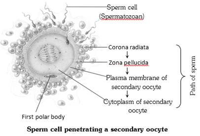 (3) Cortical reaction : Immediately after the entry of a sperm into the egg, the later shows a cortical reaction to check the entry of more sperms. In this reaction, the cortical granules present beneath the egg’s plasma membrane release chemical substance between the ooplasm and the plasma membrane (vitelline membrane).
Sperm penetration into ovum also induces following metabolic activities :
(i) The egg surface produces fertilization cone.
(ii) The vitelline membrane is lifted and is converted into fertilization membrane.
(iii) The cytoplasm exhibits movements.
(iv) The permeability of plasma membrane increases.
(v) The rate of protein synthesis increases.
(vi) Mitosis is initiated.
(4) Fusion of gametic nuclei : Entrance of spermatozoan serves to acts as stimulus which causes the second maturation division. As the head and middle piece of the sperm advance into the egg, those parts rotate through an angle of 180° so that the mitochondria and proximal centriole of the associated middle piece assume the leading position. The centriole brought in by the spermatozoan subdivides into two and as achromatic spindle is established
(3) Cortical reaction : Immediately after the entry of a sperm into the egg, the later shows a cortical reaction to check the entry of more sperms. In this reaction, the cortical granules present beneath the egg’s plasma membrane release chemical substance between the ooplasm and the plasma membrane (vitelline membrane).
Sperm penetration into ovum also induces following metabolic activities :
(i) The egg surface produces fertilization cone.
(ii) The vitelline membrane is lifted and is converted into fertilization membrane.
(iii) The cytoplasm exhibits movements.
(iv) The permeability of plasma membrane increases.
(v) The rate of protein synthesis increases.
(vi) Mitosis is initiated.
(4) Fusion of gametic nuclei : Entrance of spermatozoan serves to acts as stimulus which causes the second maturation division. As the head and middle piece of the sperm advance into the egg, those parts rotate through an angle of 180° so that the mitochondria and proximal centriole of the associated middle piece assume the leading position. The centriole brought in by the spermatozoan subdivides into two and as achromatic spindle is established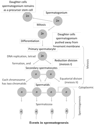 Some large cells are also found in this germinal epithelium. These are called the "Sertoli cells or Sustentacular cells". These cells provide nutrition to the maturing sperms in the form of Glycogen. For getting nutrition, the head of the sperms are submerged in the cytoplasm of sertoli cells.
Sertoli cells mainly provide nutrition and conserve the various stages of spermatogenesis. Spermatogenesis is a continuous process. To make it easier for study, it has been divided into the following steps -
(i) Formation of spermatid.
(ii) Spermiogenesis or Spermateleosis.
(i) Formation of spermatids : This process begins as the animal attains sexual maturity. The endodermal cells of the yolk sac which participate in this process are termed as the primordial germ cells. The process of formation of spermatids from primordial germ cells are termed as spermatocytosis. It has 3 sub-stages -
(a) Multiplication phase : During this process the primordial germ cells repeatedly undergo mitosis division, and as a result of these divisions spermatogonia are formed. Spermatogonia are diploid.
(b) Growth phase : Some spermatogonia either due to growth or due to food storage become 2 or 3 times of their original size, and are now known as primary spermatocytes. The remaining spermatogonia remain in the seminiferous tubules in the form of reserved stock. The primary - spermatocytes formed during the growth phase are diploid. Growth phase is the longest.
(c) Maturation phase : Primary - spermatocytes undergo Meiosis-I and as a result 2 haploid secondary spermatocytes are formed. This division is termed as First Maturation Division or Reductional division. Secondary spermatocytes undergo Meiosis II or equational division, and as result, 2 spermatids are formed from each secondary spermatocyte. Thus, from 1 diploid primary spermatocytes 2 secondary spermatocytes are formed on meiosis I and from 2 haploid secondary spermatocytes 4 spermatids are formed on meiosis-II. Metamorphosis of spermatids into sperms in known as Spermiogenesis or Spermatoliosis.
(ii) Spermatoliosis : The process of transformation of a round non-motile and haploid spermatid obtained from spermatocytosis into thread-like, motile and haploid sperm
Some large cells are also found in this germinal epithelium. These are called the "Sertoli cells or Sustentacular cells". These cells provide nutrition to the maturing sperms in the form of Glycogen. For getting nutrition, the head of the sperms are submerged in the cytoplasm of sertoli cells.
Sertoli cells mainly provide nutrition and conserve the various stages of spermatogenesis. Spermatogenesis is a continuous process. To make it easier for study, it has been divided into the following steps -
(i) Formation of spermatid.
(ii) Spermiogenesis or Spermateleosis.
(i) Formation of spermatids : This process begins as the animal attains sexual maturity. The endodermal cells of the yolk sac which participate in this process are termed as the primordial germ cells. The process of formation of spermatids from primordial germ cells are termed as spermatocytosis. It has 3 sub-stages -
(a) Multiplication phase : During this process the primordial germ cells repeatedly undergo mitosis division, and as a result of these divisions spermatogonia are formed. Spermatogonia are diploid.
(b) Growth phase : Some spermatogonia either due to growth or due to food storage become 2 or 3 times of their original size, and are now known as primary spermatocytes. The remaining spermatogonia remain in the seminiferous tubules in the form of reserved stock. The primary - spermatocytes formed during the growth phase are diploid. Growth phase is the longest.
(c) Maturation phase : Primary - spermatocytes undergo Meiosis-I and as a result 2 haploid secondary spermatocytes are formed. This division is termed as First Maturation Division or Reductional division. Secondary spermatocytes undergo Meiosis II or equational division, and as result, 2 spermatids are formed from each secondary spermatocyte. Thus, from 1 diploid primary spermatocytes 2 secondary spermatocytes are formed on meiosis I and from 2 haploid secondary spermatocytes 4 spermatids are formed on meiosis-II. Metamorphosis of spermatids into sperms in known as Spermiogenesis or Spermatoliosis.
(ii) Spermatoliosis : The process of transformation of a round non-motile and haploid spermatid obtained from spermatocytosis into thread-like, motile and haploid sperm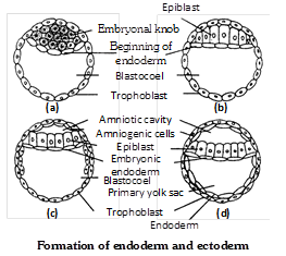
 (b) Progesterone: It is also a steriod hormone secreted by yellow-coloured endocrine gland, called corpus luteum, formed from empty Graafian folicle during the pregnancy. Small amount of progesterone is also secreted by adrenal cortex and placenta. Secretion of progesterone is


