The Fundamental Unit Of Life
Category : 9th Class
The Fundamental Unit of Life
Chapter Overview
All the living organisms which we see in our surrounding are essentially complex structures made up of numerous coordinated compartments usually known as cells. The cell is the fundamental structural and physiological unit of living organisms. Unicellular organisms consist of just one cell while multicellular organisms consist of several cells, which are specialised to perform distinct functions. A unicellular organism can perform its all metabolic activities which a multicellular organism can. The cell contains all the structures and molecular constituents needed for life.
We can compare a cell with a brick. Just as a building is made up of bricks, the body of a plant or an animal is made up of cells, i.e. all living organisms show cellular organisation, Some organisms such as Amoeba, Paramecium, Euglena, Bacteria etc. are made up of only single cell, hence are called unicellular or a cellular. There are large number of other organisms which are made up of millions of cells and are known as multicellular. All cells, whether the exist as unicellular organisms or as part of multicellular organisms demonstrate certain similar basic functions such as nutrition, respiration excretion etc. which are essential for their survival.
The history of the cell began with the invention of a microscope by the Dutch scientist Anton Van Leeuwenhoek (1632-1723) who observed the living cells of bacteria, Euglena sperms, eggs and blood corpuscles of invertebrates in 1683.
Robert Hooke (1635-1703) an English scientist, invented a primitive microscope by using lenses for achieving greater magnification. In such a microscope, the object to be seen was
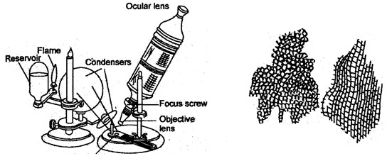
Fig.: 3.1. Primitive microscope of Robert Hooke Fig.: 5-2. Cells as seen by Robert Hooke
placed on a stage below and light coming from an oil flame was thrown on it by a convex mirror. While studying a slice of cork Robert Hooke observed a honeycomb like pattern under his microscope in 1665. He coined the term cell (cellulae), the Latin word, which means "a little room". He published his findings in Micrographic in London in 1665.
Robert Brown (1773-1858) a Scottish botanist, discovered a little sphere like structure in the cells of the orchid root in 1831. Later he named it Nucleus.
The gel like substance present in all the living cells was termed protoplasm by Hugo Von Mohl (1838) and Joharines Purkinje (1839).
Huxley (1868) called protoplasm "the physical basis of life".
Jakob Matthais Schleiden (1804-1881), a German botanist, first proposed the idea that all plants consist of cells in 1838. A year later, in 1839, a German zoologist, Theodor Schwann (1810-1882), independently asserted that all animals and plants are made up of cells. This joint finding forms the concept of cell theory. The theory states that:
Cell is the basic structural and functional unit of all living beings.
In 1855, another German biologist Rudolf Virchow gave a generalization stating "Omnis cellula-e-cellula", i.e., cell arises from a pre-existing cell.
In context of modem researches, the old cell theory has been modified and it can be stated as follows:
The function of an organism as a whole is the outcome of the combined activities and interactions of the constituent cells.
Exceptions to Cell Theory
The cells are very small structure which cannot be seen with naked eyes. In fact, human eye works as an optical instrument and the eye lens acts as a simple lens. The resolving power of a healthy young human eye is approximately 0.1 mm at 25 cm viewing distance. Any object smaller than this cannot be viewed by naked eyes. A microscope is therefore, used for enlargement of the smaller object so that it becomes visible by human eye. Microscope is a high resolution optical instrument that is used for observing fine details of very minute objects.
Compound microscope: It is also known as laboratory microscope. In this microscope many lenses are combined together and its magnification power ranges from 300 to 1500 times.
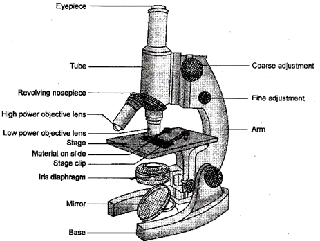
Fig: 5.1. A compound microscope
In this microscope light (usually sunlight) is used to illuminate the object, therefore, it is known as Light microscope.
The light or compound microscope is a strong, heavy metalic instrument. It comprises a U or V-shaped base having two vertical pillars. A curved arm is movably joined between the pillars to hold the microscope. The arm can be bent over the pillars of inclination joint to suit the viewer. The upper part of the arm holds the movable body tube. The body tube has eye piece and objectives. The other parts of the microscope are reflector, condenser lens, stage, eyepiece objective lens and adjustment screws (coarse and fine).
As light passes through the object, the lens nearest the object, called the objective lens, produces an enlarged image of the object in the primary image angle. The lens that you look into the eyepiece, acts as a magnifier and produces an enlarged image of the image produced by the objective lens.
To ascertain magnification, simply multiply, the eyepiece magnification, usually 10x, by the magnification of the objective lens, usually, 40x and 45x. For example a 10x eyepiece in conjugation with a 40x objective lens, will give you a magnification factor of 400. The object will be magnified 400 times larger than you can view, it with the naked eye.
Prokaryotic and Eukaryotic Cells: On the basis of complexity of structural organization .Us can be divided into two main types:
2. Eukaryotic cells
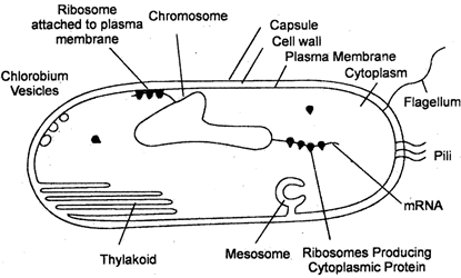
Fig. 5.2.: A typical prokaryotic cell of a bacterium
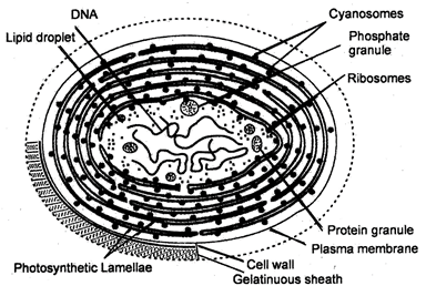
Fig. 5.3.: Photosynthetic prokaryotic cell. Ultrastructure of a cell of blue-green alga or yank bacteria
1 Prokaryotic cells (G. Pro = before; Karyon = nucleus): These cells lack a well-organised nucleis Suclear membrane around their genetic material and hence are called prokaryotic cells and the organisms which possess prokaryotic cells are known as prokaryotes.
The genetic Serial in these cells lies in direct contact with the cytoplasm and this region is called nucleiod These cells also do not contain membrane bound cell organelles like mitochondria, plastids,, endoplasmic reticulum, Golgi bodies etc. in the cytoplasm.
Ribosomes are however present 70s type. The prokaryotes include arch bacteria, bacteria and cyanobacteria (earlier called blue-green algae). The genetic material of prokaryotic cells lacks histones.
Table 5.4.Difference between Prokaryotic cells and Eukaryotic cells
|
|
Prokaryotic cells |
|
Eukaryotic cells |
|
1. |
Generally these are smaller in size |
1. |
These are generally larger in size |
|
2. |
True nucleus absent (Genetic material or nucleoid is not bounded by a nuclear membrane. |
2. |
True nucleus present (Nuclear material is bounded by a nuclear membrane). |
|
3. |
These cells contain single chromosome. |
3. |
These cells contain more than one chromosome. |
|
4. |
Chromosome lacks histones. |
4. |
Chromosomes have histones. |
|
5. |
Nucleolus is absent. |
5. |
Nucleolus is present in nucleoplasm. |
|
6. |
Membrane bounded cell organelles are absent. |
6. |
Membrane bounded organelles are present. |
|
7. |
Ribosomes are of 70s type. |
7. |
Ribosomes are of 70s and 80s types. |
|
8. |
Cell division takes place by fission or budding. |
8. |
Cell division takes place by mitotic or, meiotic cell division. |
Shape and Size of Cells
2. Cell size: A great majority of cells are too small to be seen with the naked eye but some of the cells can be seen without any optical instrument (e.g. egg of birds, reptiles and some insects, jute fibres etc.). The size of cells vary from the small cells of bacteria (0.2 to\[5.0\mu m\]) to the very large egg of the ostrich (18 cm). Mycoplasma (Pleuropneumonia like
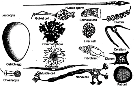
Fig. 5.5.: Various types of eukaryotic cells showing different shapes
organism; PPLO) is smallest sized cell (0.1 to \[5.0\mu m\]) while a nerve cell is longest animal cell (about 90 to 100 cm). Acetabularia, a single celled marine alga measures nearly 10 cm in height. The fiber cells (e.g. Sclerenchyma cells) of manila hemp similarly are over 100 cm in length.
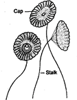
Fig. 5.6 Acetabularia
Measurement of Cell
The cell is generally measured in micron\[(\mu )\]. Micron is a unit of length in the CGS system, equal to one millionth of a meter. In SI units it is replaced by the micrometer\[(\mu m)\].
1 Micrometer \[={{10}^{-6}}\] meter
1 Millimeter \[={{10}^{-3}}\mu \]or \[1000\mu \]
1 Nanometer = Mill micron \[=1000m\mu \]
1 Angstrom \[\left( \overset{\circ }{\mathop{A}}\, \right)={{10}^{-10}}\,meter\,\,={{10}^{-7}}\,\,mm\]
Do You Now
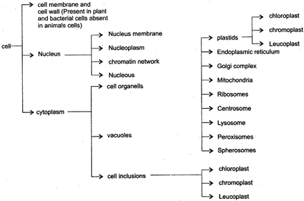
Although, all cells from different sources do not look similar in their shape, size and activities, but structurally all eukaryotic cells have three main components:
Plasma membrane is the outermost boundary of cell, but in plant cell it is also surrounded outwardly by a rigid structure. Cell wall. Inside the plasma membrane various cell organelles and inclusions are suspended in the cytoplasm including nucleus, though nucleus is separated from cytoplasm by a membrane known as nuclear membrane.
Chemical analysis has shown the membrane to be 75 percent phospholipids. In addition, i.e. membrane possesses proteins, cholesterol, and polysaccharides. However, these are the phospholipids that form key elements in the structure of plasma membrane.
Kinds of Permeability
(i) Impermeable: If a membrane does not allow to pass through it both solvent and solute molecules, it is known as impermeable membrane, e.g. cuticle layer.
(ii) Semi-permeable: If a membrane allows to pass only solvent molecules but not solute) articles through it, it is called semipermeable membrane, e.g. artificial vapour membrane.
(iii) Permeable: If a membrane allows to pass both solvent and solute molecules through it freely, it is called permeable membrane, e.g. primary cell wall.
(iv) Selectively permeable: If a membrane allows penetration of solvent freely but selects the passage of specific solute particles, it is known as selectively permeable membrane, e.g. plasma membrane.
Functi0ons of Plasma Membrane
The primary function of the plasma membrane is the regulation of various substances in and out of the cell. Some of the important functions of the cell membrane are given below:
Transport across Cell membrane
The plasma membrane acts as a barrier for the free movement of ions and metabolites across the cell, but at the same time it allows the transport of selective metabolites across it and thus regulates the flow of meterials into and out of the cell. Transport of material across the membrane is of two types:
(a) Diffusion
(b) Osmosis
(a) Diffusion: It is the transport of materials (solids, gases or liquids) from a region of higher concentration to a region of lower concentration, so as to spread uniformly in the given space.
Thus, the process of diffusion continues as long as the substance in the form of molecules or ions is not uniformly diffused. The diffusion of a substance is independent of the presence of other diffusing substances.
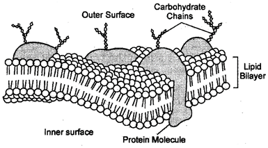
Fig. 7.1: Plasma membrane
Importance of diffusion:
(i) Gaseous exchange: Diffusion helps in gaseous exchange between the cells and the environment.
(ii) Osmosis: It is a type of diffusion where only solvent is allowed to diffuse.
(iii) Intracellular Distribution: It helps in spread of various substances throughout cytoplasm of cell.
(iv) Transpiration: Loss of water in vapour form from the aerial parts of the plant take place through diffusion.
(v) Aroma: Flowers and fruits spread aroma through diffusion to attract insects, bin and other animals for pollination and dispersal.
(b) Osmosis: The process of transport of water or any other solvent molecules from a region of lower concentration to a region of higher concentration through a semi-permeable membrane is known as osmosis. In other words "Osmosis is the process in which water molecules moves from its higher concentration to its lower concentration through semi- permeable membrane."
Osmosis is purely a mechanical diffusion process by which cell or root hair absorb water without spending any amount of energy.
Demonstration of Osmosis
Take a thistle funnel and cover its mouth with a piece of an artificial semipermeable membrane like parchment paper or animal bladder. Invert the thistle funnel and place it into a beaker filled with dean and pure water. Pour concentrated salt or sugar solution into thistle funnel till it reaches one third of the height of stem. Fix the stem of thistle funnel in a vertical position by means of a stand. Make a mark at the level of solution as 'A’ with the help of a glass marking pencil.
Few hours later, a rise in the level of solution in the thistle funnel tube will be observed. Mark this level as 'B’ by means of glass marking pencil or marker.
The concentration of water molecules in beaker is higher and these water molecules are passed through the semipermeable membrane, because the sugar solution filled in the thistle funnel has lower water concentration. Thus there is an increase in the level of solution in the thistle funnel. With the gradual increase in the level of water inside the thistle funnel the concentration of sugar solution will decrease and gradually the diffusion of water molecules into thistle funnel decreases.
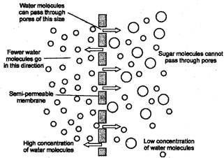
Fig. 7.2: Movement of molecules through a semipermeable membrane
Osmotic solutions are those solutions which can cause osmosis if separated from solvent by means of a semipermeable membrane. These solutions are of three types:
(a) Hypotonic Solutions: These are dilute solutions which have an osmotic concentration lower than that of another solutions.
(b) Isotonic Solutions: These solutions have an osmotic concentration similar to that of another solutions.
(c) Hypertonic Solutions: These are concentrated solutions which have an osmotic concentration higher than that of another solutions.
Types of Osmosis
On the basis of concentration of cell and its external solution osmosis is of two types:
(a) Endosmosis: It is the osmotic entry of water in to a cell.
(b) Exosmosis: It is the osmotic withdrawal of water from a cell.
Importance of Osmosis
(i) Absorption: Osmosis helps in absorption of water and minerals from the soil by root hair.
(ii) Turgidity: It helps in developing turgor pressure which helps in opening and closing of stomata and movements of leaflets in Mimosa pumice.
(iii) Cell to cell water movement: Cells absorb or loose water to one another through osmosis.
(iv) Seed germination: It helps in growth of radicle and plumule during seed germination.
(v) It maintains turgidity of cells which helps in deeper penetration of roots, extension of leaves, stretching of stem etc.
These are energy dependent processes so are rapid, energy is provided by hydrolysis of ATP. By this process materials can be transported completely across the cell membrane. It is highly affected by low temperature, absence of oxygen and presence of donator phenol, cyanides etc.
Glucose, amino acids and some ions pass through the plasma membrane by an active transport or transport.
(a) Endocytosis: It is the process by which large sized liquid or solid particles are taken inside the cell through plasma membrane. It is further divided into two types-
(i) Pinocytosis: It is also known as cell drinking. It is the process which involves intake of large sized liquid nutrients, while the cell showing pinocytosis, and is called pinocyte. It Extracellular Fluid Extracellular Fluid was first observed in Amoeba. In this, the extra cellular solutes are taken into a pocket formed by invagination of plasma membrane and called pinocytotic vesicle. It later separates from the cell membrane as sin infra cellular food vacuole, called pinosome. Pinosome is digested by the hydrolytic enzyme of primary lysosome.
(ii) Phagocytosis: It is known as cell- eating. It is the process which involves the intake of large-sized solid particles including cellular-debris and microbs.
Phagocytosis is the major feeding method in many unicellular organisms (e.g Amoeba), simple metazoa (e.g. Sponges) and WBC and macrophage cells also. In this, the material to be ingested first binds to the cell surface which invigilates to form a phagocytotic vesicle. It later separates from cell membrane as an intracellular food vacuole, called phago-some.
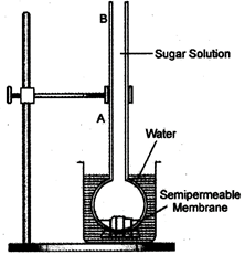
Fig. 7.3: Experiment to demonstrate the process of osmosis by means of thistle funnel
Osmotic solutions are those solutions which can cause osmosis if separated from solvent by means of a semipermeable membrane. These solutions are of three types:
(a) Hypotonic Solutions: These are dilute solutions which have an osmotic concentration lower than that of another solutions.
(b) Isotonic Solutions: These solutions have an osmotic concentration similar to that of another solutions.
(c) Hypertonic Solutions: These are concentrated solutions which have an osmotic concentration higher than that of another solutions.
Types of Osmosis
On the basis of concentration of cell and its external solution osmosis is of two types:
(a) Endosmosis: It is the osmotic entry of water in to a cell.
(b) Exosmosis: It is the osmotic withdrawal of water from a cell.
Importance of Osmosis
(i) Absorption: Osmosis helps in absorption of water and minerals from the soil by root hair.
(ii) Turgidity: It helps in developing turgor pressure which helps in opening and closing of stomata and movements of leaflets in Mimosa pumice.
(iii) Cell to cell water movement: Cells absorb or loose water to one another through osmosis.
(iv) Seed germination: It helps in growth of radicle and plumule during seed germination.
(v) It maintains turgidity of cells which helps in deeper penetration of roots, extension of leaves, stretching of stem etc.
These are energy dependent processes so are rapid, energy is provided by hydrolysis of ATP. By this process materials can be transported completely across the cell membrane. It is highly affected by low temperature, absence of oxygen and presence of donator phenol, cyanides etc.
Glucose, amino acids and some ions pass through the plasma membrane by an active transport or transport.
(a) Endocytosis: It is the process by which large sized liquid or solid particles are taken inside the cell through plasma membrane. It is further divided into two types-
(i) Pinocytosis: It is also known as cell drinking. It is the process which involves intake of large sized liquid nutrients, while the cell showing pinocytosis, and is called pinocyte. It Extracellular Fluid Extracellular Fluid was first observed in Amoeba. In this, the extra cellular solutes are taken into a pocket formed by invagination of plasma membrane and called pinocytotic vesicle. It later separates from the cell membrane as sin infra cellular food vacuole, called pinosome. Pinosome is digested by the hydrolytic enzyme of primary lysosome.
(ii) Phagocytosis: It is known as cell- eating. It is the process which involves the intake of large-sized solid particles including cellular-debris and microbs.
Phagocytosis is the major feeding method in many unicellular organisms (e.g Amoeba), simple metazoa (e.g. Sponges) and WBC and macrophage cells also. In this, the material to be ingested first binds to the cell surface which invigilates to form a phagocytotic vesicle. It later separates from cell membrane as an intracellular food vacuole, called phago-some.
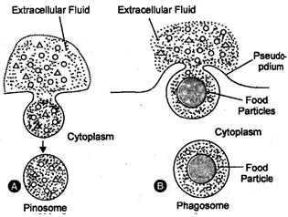
Fig. 9.1. A. Pinocytosis B. Phagocytosis
(b) Exocytosis-It is also called emeiocytosis or cell vomiting or ephagy. It involves the expelling of large sized particles outside the cell through cell membrane.
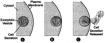
Fig.9.2.: Exocytosis by a cell
In this process, the materials to be expelled outside the cell, are accumulated in a membrane round vesicle, called exocytosis vesicle, which moves outwards, fuses with plasma membrane 3ind the contents are discharged outside the cell. Exocytosis is responsible for-
(i) removal of undigested food left in the food vacuoles in the cells.
(ii) replacement of internalized membrane by the fusion of exocytosis vesicle with the cell membrane.
Cell wall is the outer most, rigid, protective and supportive covering found in all the plant cells, bacteria, cyanobacteria and some protists. It is absent in animal cells. It was first discovered by Robert Hooke (1665 AD).
Structurally, the plant cell wall is formed of two coats-
(i) Primary cell wall: It is outer, thinner, elastic coat and is mainly made of cellulose.
(ii) Secondary Cell wall: It lies inner to primary cell wall only in mature and non-dividing cells.
It is chemically formed of cellulose, hemicellulose and further deposited with suberin or lignin.
Cellulose is a complex fibrous carbohydrate (homo polysaccharide) which cannot be digested by human beings and several other animals. It is most abundant organic molecule. Cell wall of fungi is made up of chitin.
Functions of Cell Wall
(1) The cell wall provides protection to inner contents from mechanical injuries and entry of germs.
(2) It provides mechanical support (due to rigid cell walls) to the aerial parts.
(3) It maintains shape of the cell.
(4) Cell wall of root hair helps in absorption of water and minerals from the soil.
(5) It prevents undue expansion of the cell.
(6) Suberin and cutin present in cell wall prevent evaporation of water.
(7) It helps in keeping a balance between intracellular osmotic pressure and that of environment.
The nucleus was first observed by Robert Brown (1831 A. D.) in orchid cells. It is the most conspicuous and the largest organelle of a eukaryotic cell. It is visible under light microscope and its fine structure has been revealed by electron microscope. The nucleus can be called "controlling centre of the cell" since it contains chromosomes and genes, which control all activities of an individual cell.
Occurrence: A true nucleus is present in all the eukaryotic cells except mature mammalian RBCs, sieve-tube cells of phloem. The prokaryotic cells have an incipient nucleus, called nucleoid or genophore.
Position: The nucleus is generally centric but is peripheral in adipocytes and basal in the columnar and gland cells. In Spirogyra, it is suspended in the centre by the cytoplasmic strands while in most of plant cells, it is extrinsic due to presence of large central vacuole.
Number: Generally, there is a single nucleus in each cell. Such cells are known as uninucleate. Some cells contain two nuclei and are known as binucleate(e.g. Paramecium Caudatum).
Some cells possess more than two nuclei. They are termed Multinucleate (e.g. Vaucheria).
Shape and Size: The shape and size of the nucleus varies with the type and function of the cell. The shape of nucleus is generally spherical cuboidal (germ cells), oval (columnar cells of intestine), discoidal (flat cells of endothelium), kidney shaped (meganucleus of Paramecium horse-shoe shaped (meganucleus of varticella), bilobed (acidophils), 3-lobed (basophils), multiplied (neutrophils), long and mpruliform (stentor) and branched (labyrinthine).
Chemical Composition: Chemically, the nucleus is composed of about 80% proteins (65% acidic and 15% basic), 12% DNA, 5% RNA and 3% lipids
Structure: The nucleus is composed of following components:
(i) Nuclear envelop or Karyotheca
(ii) Nuclear sap or Karyolymph
(iii) Chromatin material
(iv) Nucleolus and
(v) Nuclear matrix
(i) Nuclear Envelop: Nuclear envelop or Karyotheca is a double membranous covering which separates the nucleus from the cytoplasm. Nuclear pores on these two membrane allow exchange of materials between the nucleus and cytoplasm. The outer membrane of nuclear envelop is usually connected with endoplasmic reticulum (ER). It also bears ribosomes.
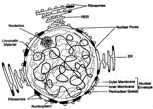
Fig. 11.1: Structure of nucleus
(ii) Nucleoplasm or Nuclear Sap: It is transparent, homogenous, semifluid, colloidal ground substance present inside the nuclear membrane, in which chromatin and nucleolus are embedded It is chemically formed of water, sugars minerals, nucleotides, ribosomes, enzymes, m-RNA and + RNA molecules.
(iii) Nucleolus: It was first observed by Fontana. It is nearly spherical structure found inside the nucleus. In certain cases, a nucleus may have two or more nucleolus. Nucleolus not bounded by a membrane. It is rich in protein and RNA. It is the site of synthesis of ribosome Ribosomes are helpful in protein synthesis in cytoplasm.
(iv) Chromatin Material: The chromatin material is thin, thread like-intertwined substance composed of the genetic substance DNA (deoxyribo nucleic acid) and proteins (Histone and histone). Chromatin also contains some RNA, certain metallic ions and some enzymes. During cell division, chromatin becomes highly condensed, thick, rod like structure known as Chromosomes.
Chromosomes: During cell division, the chromatin fibres condense by spiralisation and dehydration into a number of rod like structures, called chromosomes. Chromosomes contain hereditary in formations of the cell in the form of genes.
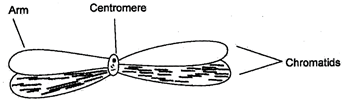
Fig. 11.2. A Chromosome
Functions of Nucleus
The nucleus performs following functions:
Cytoplasm is an amorphous colloidal ground substance lying between the plasma membrane and the nucleus. It contains various organelles and cell inclusions. Often under the free surface of the cell, the cytoplasm is more viscous and called ectoplasm. The internal cytoplasm is, however, less-viscous and is known as endoplasm.
Cytoplasm is differentiated into three parts:
(i) Cytosol or Cytoplasmic matrix,
(ii) Cell organelles and
(iii) Cell inclusions
The liquid and soluble part of cytoplasm is nearly transparent and is called cytosol, or cytoplasmic matrix or hyaloplasm. It occurs between the cell organelles. Cytosol contains a system of protein fiberes called cytoskleton, but otherwise appears transparent and structures in the electron microscope. It contains about 90% of water, various cell organelles and inclusions in it. Cytosol also contain ions and biomolecules such as sugars, nucleotides, amino acids, minerals, enzymes, vitamins, dissolved gases etc. These are necessary for the biosynthetic, processes of the cell. The cytosol contains waste materials too. All these substances may be present either as true solutions or as colloids.
Functions:
(i) It provides raw materials to various cell organelles for their functioning.
(ii) It helps in exchange of material between various cell organelles and also with ECF.
(iii) If is the seat of biosynthesis of organic biomolecules like fats, nucleotides, proteins etc.
(iv) It distributes the nutrients, metabolites, enzymes and other substances in the cell.
(b) Streaming of cytoplasmic matrix (cylosis) serves many functions in the cell.
Large and complex cells, including cells from multicellular organisms, need a lot of chemical activities to support their complicated structure and function. To keep these activities of different types separate from each other, these cells need compartmentalization and use double membranous little structures or cell organelles within themselves. This is one of characteristic of the eukaryotic cells that distinguish them from prokaryotic cells. Some of these organells are visible only under the electron microscope. Except plasma membrane and cell wall, all other cell organelles are found embedded in the cytosol.
We have discussed about the nucleus in a previous section. Some important examples of cell organelles which we discuss now are; endoplasmic reticulum, Golgi apparatus, lysosomes, mitochondria, plastids and vacuoles, They are important because they carry out some very crucial functions in the cells.
Table 14.1: Differences between organs and organelles
|
|
Organs |
|
Organelles |
|
1. |
They are found in only multicellular organisms. |
1. |
They are found in all eukaryotic cells. |
|
2. |
Organs are large sized or microscopic. |
2. |
These are very small sized, either microscopic or sub microscopic. |
|
3. |
They may be external or internal. |
3. |
They are always in ternal. |
|
4. |
The organ is formed of tissues, tissues of cells and cells of organelles. |
4. |
An organelle is form of micro molecules and macromolecules. |
|
5. |
Organs coordinate to form organ system, while organ system form the body of organism. |
5. |
Organelles coordinate to produce cells. |
Endoplasmic reticulum (ER) was discovered independently by Porter (1945) and Thompson (1945) and was named by Porter in 1953. It is absent in the prokaryotic cells but is present in all the eukaryotic cells except germinal cells and mature mammalian erythrocytes, (RBCs). The- striated muscle fibres have a special type of ER called Sarcoplasmic reticulum (SR).
Endoplasmic reticulum is a complex network of membrane-bound channels or sheets, tubules and vesicles. It may be connected to both the plasma membrane as well as outer nuclear membrane. The membranes of endoplasmic reticulum are similar in structure to the plasma membrane.
Structure: ER consists of membrane lined channels or spaces which have three characteristic forms cisternae tubules and vesicles.
(i) Cisternae are flattened, unbranched, sack like elements with a diameter of 40 to 50 nm.
They lie in stalks parallel to one another.
(ii) Tubules are tube like extensions which may be connected with cisternae or vesicles to form a reticular system.
(iii) Vesicles are oval or rounded, vacuole like elements. They often occur isolated in the cytoplasmic matrix.
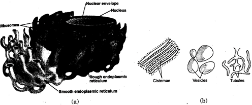
Fig.15.1: (a) Endoplasmic Reticulum (b) Various parts of ER
Table 15.2: Differences between smooth ER and Rough ER
|
|
Smooth Endoplasmic Reticulum (SER) |
|
Rough Endoplasmic Reticulum (RER) |
|
(i) |
It consists mainly of tubules and vesicles. |
(i) |
It mainly consists of cisternae. |
|
(ii) |
It is free of ribosomes. |
(ii) |
I It has ribosomes on its cytoplasmic |
|
(iii) |
It is specialized to synthesize lipids and steroids. |
(iii) |
It is specialized to synthesize proteins. |
|
(iv) |
The products do not pass into lumen, |
(iv) |
The products pass into lumen of ER for transport to other places. |
Functions of ER
(i) ER is a component of cytoplasmic-vascular system which acts as structural framework and provides mechanical support and shape to the cell.
(ii) ER acts as cell circulatory system and helps in transportation of materials in a directional flow.
RER \[\to \]SER \[\to \]Golgi body \[\to \]Primary lysosome out of cell.
(iii) Rough ER is the site of protein synthesis.
(iv) Smooth ER helps in lipid synthesis.
(v) RER also helps in the synthesis of nuclear envelop during telophase of cell division.
(vi) It helps in storage of synthetic products like glycogen.
(vii) SER helps in glycogenolysis in the liver cells.
(viii) In releases \[C{{a}^{++}}\]ions required for muscle contraction.
Golgi apparatus, also called Lipochondria, or Golgi body or Golgi complex, was discovered by Camilo Golgi in 1898 in the nerve cells of cats. It is present in all eukaryotic cells except mature red blood cells and sperms of mammals and absent in prokaryotic cells, In the vertebrate cells Golgi body is single and localized and occupies a fixed position. It lies around or above centrioles near the nucleus, while a number of Golgi bodies are pronuclear in the nerve cells. In the invertebrates and plant cells, a number of Golgi bodies are scattered in the cytoplasm and are called dictyosomes.
Structure: The Golgi body is formed of three types of components.
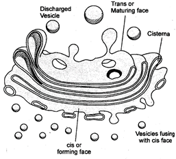
Fig.16.1 Golgi apparatus
(a) Cisternae: These are elongated double membranous, flat and slightly curved components with swollen ends present one above the other, also called flattened sacs or macules. These are about 3-12 in animal cells and 10-20 in plant cells and certain protists.
In fungi, a dictyosome is unicisternal.
(b) Vacuole: These are spherical components and lie towards the concave side of the cisternae.
(c) Vesicles: These are small sized components present along the convex surface or edges of the cisternae. Vesicles are of two types-Smooth vesicles, which contain secretory products of ER and Golgi body and Coated Vesicles with rough surface and generally lie near the convex surface.
The Golgi body has a definite polarity. Its concave side is always directed towards the cell membrane and is called maturation face or trans face while its convex surface is directed towards the nucleus and is called formative face or cis face. It appears that materials are transported from cis to trans face by vesicles.
Origin: Most accepted view is that Golgi apparatus orginates from endoplasmic reticulum.
It is supported by the fact that cisternae of Golgi apparatus are continuous with SER at certain places.
Functions:
Lysosomes were first discovered by Belgian cytologist and biochemist Christoin de Duve in 1949. The term was coined by Novikoff and derived from two greek words: lysis = digestive, soma = body.
Lysosomes are electron microscopic, vesicular structures of the cytoplasm which are involved in intra-cellular digestive activities, so known as lysosomes.
Occurrence: Lysosomes are present in all eukaryotic animal cells (except mammalian RBCs), some fungi, some protists and meristem tic cells (root tip cells of maize) in plant cells. These are absent in prokaryotic
Structure: Lysosomes are tiny, globular structures evenly scattered in the cytoplasm. Each lysosome is bounded by a single unit membrane made up of lipoprotein. It contains a dense, finely granular fluid consists of hydroly tic enzymes. These hydrolytic enzymes are capable of digesting most
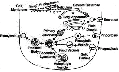
Fig.17.1: Formation of lysosomes and intracellular of organic matter, digestion in them
Fig.17.1: Formation of lysosomes and intracellular of organic matter, digestion in them
Lysosomes are of four types:
(i) Primary lysosomes have only digestive enzymes in inactive form and are also called storage granules.
(ii) Secondary lysosomes have digestive enzymes and ingested food. It is formed by fusion of primary lysosomes and endosomes.
(iii) Auto phagosomes are formed of primary lysosome and some cell organelles, during deficiency of foreign food.
(iv) Residual body or tertiary lysosome is the lysosome with only undigested food.
The Lysosomes may be called "suicide bags" of the cell in view of their autolytic role, or "disposal unit" of the cells because they remove the foreign bodies, toxic molecules, and debris; or "recycling centres" as they break down worn out cells and cell organelles.
Functions:
(1) Lysosomes help in intracellular digestion of food particles.
(2) They help in the destruction of foreign particles, such as bacteria and viruses so they provide protection to the body.
(3) They help in removing cell debris, dead and worn out cellular organelles by digesting them. Therefore called Cellular scavengers.
(4) During metamorphosis in several animals, the larval organs are digested by lysosomes to provide raw materials for the formation of adult organs.
Mitochondria are small sausage or rod-shaped bodies, which are called the "power house of the cell". They are associated with cellular respiration and are the sources of energy.
These were first discovered by Kolliker in 1880 in the voluntary muscles of flying insects.
Term mitochondria (mito = thread; Chondrion = granule) was given by C. Benda in 1897.
Occurrence: These are found in all eukaryotic cells except mature and old mammalian RBCs. These are absent in prokaryotic cells.
Shape: The shape may be, fibrillar, spherical (in yeast), oval, sausage shaped or discoidal.
Number and Position: The number of mitochondria depends upon the metabolic state of the cell. They are more in growing, dividing and metabolically active cells.
Minimum number if it is one in yeast, few in green algae and protozoans. Maximum number of mitochondria (500000' per cell) are found in flying muscle cell of insect.
The mitochondria are generally located in metabolically active areas of the cells to provide ATP immediately for quick cellular activities.
Structure: Mitochondria are semiautonomous organelles bound by an envelope of two unit membranes and filled with a fluid matrix. The outer membrane is smooth and has porous proteins which forms channels for the passage molecule through it. The inner membrane is semipermeable. It usually produces numerous in folds called cristae.
Fig. 18.1: A longitudinally cut mitochondrion
The cristae greatly increase the inner surface area of the mitochondria to hold a variety of enzymes. Cristae and inner membrane bear minute, regularly spaced tennis racket shaped wide particles known as\[{{F}_{1}}\] particles or oxysomes. The membrane of oxysomes have various respiratory enzymes. Cristae increase the surface area for ATP generating chemical reactions.
ATP synthesis occurs over \[{{F}_{1}}\]particles. Mitochondrial membrane encloses a matrix having DNA, ribosomes and enzymes. The DNA and ribosomes make the mitochondria semi-autonomous as they are able to manufacture some of their own proteins and enzymes.
Do You Know
Why mitochondria are termed semi-autonomous?
Mitochondria are called semi-autonomous organelles because of the following reasons:
Functions:
The term plastid was introduced by E. Haeckel in 1866. Plastids are semiautonomous organelles having DNA and double membrane envelop which store or synthesize various types of organic compounds.
Location: Plastids are found in the plant cells and in certain protists but are absent to animal eels.
Types: According to their structure, pigments and functions, plastids are of three types:
Structure: The Chloroplasts of higher plants are usually spherical, ovoid, and discoid or lens- shaped. Each chloroplast is bounded by an envelope of two unit membranes and is filled with a fluid matrix like a mitochondrion. It consists of three parts-envelop, matrix and thylakoids.
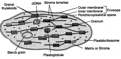
Fig.19.1: Internal structure of chloroplast
Matrix is colourless, granular, colloidal ground substance called stromal. It contains proteins, lipids, 70s ribosomes, a small circular double helical DNA molecule, RNA molecules, enzymes, lipid droplets called plastoglobuli and certain metal ions.
Inside the matrix there is a series of parallel membranous sheets called lamellae, which form a number of oval-shaped closed sacs, called thylakoids. Thylakoids are structural and functional elements of Chloroplasts. Thylakoids contain all the requirements of light reactions eg. chlorophyll, carotenoids, plastoquinone, plastocyanin etc. In eukaryotic plant cells, a number of thylakoids are superimposed like a pile of coins to form a granum (plural = grana).
Granum is the site of light reaction during photosynthesis where as stroma is the site of dark reaction during photosynthesis.
Semi-autonomous nature of chlorplast: Like the mitochondria, the chloroplast are semi- autonomous organelles because these have complete machinery (e.g. DNA, RNA, ribosomes and ATP) to synthesize some of the required proteins while for some other proteins these depend upon nuclear DNA and cytoplasmic ribosomes.
Functions:
(i) The Chloroplasts trap the radiant energy of sunlight and transform it into the chemical energy of carbohydrates using water and \[C{{O}_{2}}\] in the presence of chlorophyll. Thus these are called kitchen of cell. This process is called photosynthesis.
(ii) These maintain oxygen/carbon dioxide balance in the biosphere.
(iii) These store the starch in the protenacious bodies called pyrenoids in algal forms.
They are found in flowers and fruits and provide them characteristic colours. They contain the pigments carotenoids which include carotenes and xanthophylls.
The chromaplasts impart various colours to flowers to attract insects for pollination and to the fruits for alluring certain animals for seed dispersal.
Leucoplasts store food in the form of starch, fats and proteins. Thus these are called store house of the cell.
Table 19.2: Differences between chloroplast and chromoplast
|
Chlosoplast |
Chromoplast |
|
1. They are green in colour. |
1. They are coloured (except green colour) |
|
2. They have chlorophyll and carotenoids. |
2. Chlorophyll are absent, only carotenoids are |
|
3 . Lamellae are present in the chloroplast. |
3. Lamellae are absent. |
|
4. Chloroplasts are site of photosynthesis. |
4. They add colour to the flowers, fruits, for attracting pollinators and dispersal agents. |
Vacuoles are storage sacs for solid or liquid contents. These are found in all the eukaryotic cells. In animal cells, there are a number of small sized vacuoles, irregularly scattered in the cytoplasm and are generally in modified form. On the other hand plant cells, have one or two very large vacuoles. The central single vacuole of some plant cells may occupy 50-90% of the cell volume.
Structure-A vacuole is formed of two parts-
(a) Tonoplast-It is a outer, single layered, unit membrane and is semipermeable.
(b) Cell sap-It is a liquid filled inside the central vacuole and is formed of water, minerals, sugars, amino acids, proteins, esters, excretory substances, alkaloids, organic acids etc.
The vacuoles are modified differently to perform different functions-
(i) Food vacuoles-These possess ingested or digested or undigested food and may be in the form of pinosome or phagosome. These are found in phagocytes and protozoans (e.g. Amoeba).
(ii) Contractile Vacuole-These are found in fresh water protozoans e.g. Amoeba, Paramecium etc. and help in osmoregulation (regulating water contents inside cell) and excretion.
(iii) Gas or air vacuoles (Pseudo vacuoles)-These are found in cyanobacteria and provide buoyancy as they contain the metabolic gases.
Functions:
(i) Cell sap exerts turgor pressure which keeps the plant cell turgid.
(ii) It stores water, nutrients and minerals.
(iii) Vacuoles act as dump house of excretory waste products in plant cells.
(iv) Contractile vacuoles take part in osmoregulation and excretion.
(v) Gas vacuoles provide buoyancy in some plant cells.
Ribosomes were discovered by Robinson and Brown (1953) in plant cell and by Palade (1955) in animal cell. Palade also coined the term Ribosome.
Ribosomes are smallest known electron microscopic, ribonucleic-protein particles attached either on RER or floating freely in the cytoplasm and are the site of protein synthesis.
Occurrence: These are found in both prokaryotes as well as eukaryotes except mature RBCs and sperms. In the prokaryotes these are present only in free form in the cytoplasm while in eukaryotes these are found on RER or free form.
Types: On the basis of sedimentation co-efficient, ribosomes are of two types-
(a) 80s Ribosomes: These are found in eukaryotic cells. Each 80s ribosome consists of a large 60s sub-unit and a small 40s sub-unit.
(b) 70s Ribosomes: These are found in prokaryotic cells and in the plastids of eukaryotic cells. Each 70s ribosome consists of a large 50s submit and a small 30s sub-unit.
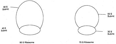
Fig. 21.1
Function: Ribosomes are sites for protein synthesis in the cell, hence they are popularly known as protein factories of the cell. They provide site for attachment of tRNA and mRNA which participate in protein synthesis.
Peroxisomes
These were discovered by Rhodin in liver and kidney cells. These are found in photosynthetic cells of the plants, and liver and Kidney cells of vertebrates.
These are spherical in shape, about \[1-5\mu m\]in size. Each contains two types of enzymes bounded by a unit membrane.
(i) Oxidases: These oxidise organic compounds.
(ii) Catalases: These degrade \[{{H}_{2}}{{O}_{2}}\]
Function: are involved in the formation and degradation of\[{{H}_{2}}{{O}_{2}}\]. Plant peroxisomes are also involved in photorespiration.
Centrosomes
Centrosome is found only in the animal cells. It is not surrounded by any membrane but consists of two granule like centroiles. The centrioles commonly pairs, a pair of centrioles is called a diplosome.
A centriole possesses a whorl of nine peripheral fibrils.
Fibrils are absent in the centre. The centre of centriole contains a rod-shaped proteinaceous mass known as hub.
From the hub, arises 9 strands towards the peripheral triplet fibrils called radial fibres of spokes.
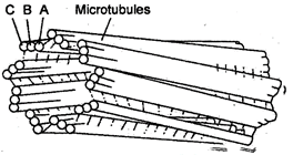
Fig.21.2 Centriole
Functions
(i) The centrioles form asters during cell division of animal cells while in the dividing plant cells, the spindle is anastral.
(ii) Distal centriole of sperm gives rise to axoneme of sperm-tail.
Chapter at a Glance and Glossary


You need to login to perform this action.
You will be redirected in
3 sec

