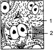
A) Figure - Cartilage 1- Collagen 2- Chondrocyte
B) Figure - Cartilage 1- Collagen 2- Chondroclast
C) Figure - Bone 1- Microtubule 2- Chondroclast
D) Figure - Bone 1- Collagen fibres 2- Osteoblast
Correct Answer: A
Solution :
[a] The given figure represents the image of cartilage. Cartilage is an important structural component of the body. It is a firm tissue but is softer and much more flexible than bone It is a connective tissue found in joints between bones e.g. the elbows, knees and ankles- ends of the ribs; between the vertebrae in the spine; ears and nose; bronchial tubes or airways. Cartilage is made up of specialised cells called chondrocytes (2) These chondrocytes produce large amounts of extracellular matrix composed of collagen fibres, proteoglycan, and elastin fibres There are no blood vessels in cartilage to supply the chondrocytes with nutrients.


You need to login to perform this action.
You will be redirected in
3 sec

