Muscular and Skeletal System
Category : UPSC
Muscular and Skeletal System
Skeletal Systems of Various Animals
(i) Movement is a major characteristic of animals. This movements is a result of contraction of muscles. The skeleton helps transmit that movement. Skeletons are either a fluid-filled body cavity, exoskeletons or internal skeletons.
(ii) Hydrostatic skeletons consist of fluid-filled closed chambers. Internal pressures generated by muscles contraction cause movement as well as maintain the shape of the animals, such as the sea anemone and worms. The sea anemone has one set of longitudinal muscles in the outer layer of the body, and a layer of circular muscles in the inner layer of the body. The anemone can elongate or contract its body by contracting one or the other set of muscles.
(iii) Exoskeletons are characteristic of the Phylum Arthropoda. Exoskeletons are hard segments that cover the muscles and visceral organs. Muscles for movement attach to the inner surface of the exoskeleton
Exoskeletons restrict the growth of the animal, thus it must shed its exoskeleton (or molt) to form a new one that has room for growth. The bulk and weight of the exoskeleton and associated mechanical attain.
Note: Spiders use a combination of an exoskeleton for protection and fluid pressure for movement.
Vertebrates have developed an internal mineralized (in most cases) endoskeleton composed of bone and/or cartilage. Muscles are on the outside of the endoskeleton.
Cartilage and bone are types of connective tissue.
- Sharks, and rays have skeletons composed entirely of cartilage; other vertebrates have an embryonic cartilage skeleton progressively replaced by bone as they mature and develop.
- Some areas of the human body, however, retain cartilage in the adult: in joints and flexible structures such as the ribs, trachea, nose and ears.
The Skeleton and Muscles
- The skeleton and muscles function together as the musculoskeletal system. This system (often treated as two separate systems, the muscular, and skeletal) plays an important homeostatic role: allowing the animal to move to move favorable external conditions.
- Certain cell in the bones produce immune cells as well as important cellular components of the blood.
- Bone also helps regulate blood calcium levels, serving as a calcium sink. Rapid muscular contraction is important in generating internal heat, another homeostatic function.
Types of Skeletons
- The axial skeleton consists of the skull, vertebral column, and rib cage.
- The appendicular skeleton contains the bones of the appendages (limbs, wings, or flippers/fins), and the pectoral and pelvic girdles.
- The human skull, or cranium, has a number of individual bones tightly fitted together at immovable joints.
- At birth many of these joints are not completely structured together as bone, leading to a number of “soft spots” or fontanels, which do not completely join until the age of 14-18 months.
- The vertebral column has 33 individual vertebrae separated from each other by a cartilage disk. These disks allow a certain flexibility to the spinal column, although the disks deteriorate with age, producing back pain. The sternum is connected to all the ribs except the lower pair. Cartilage allows for the flexibility of the rib cage during breathing.
- The arms and legs are part of the appendicular skeleton.
- The upper bones of the limbs are single: humorous (arm) and fernur (leg).
- Below a joint (elbow or knee), both limbs have a pair of bones (radius and ulna in the arms; tibia and fibula in legs) that connect to another joint (wrist or ankle).
- Each hand or foot ends in 5 digits (fingers or toes) composed of metacarpals (hands) or metatarsals (feet).
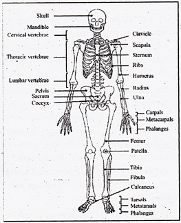
- Limbs are connected to the rest of the skeleton by collections of bones known as girdles. The pectoral girdle consists of the clavicle (collar bone) and scapula (shoulder blade).
- The humerus is joined to the pectoral girdle at a joint and is held in place by muscles and ligaments. A dislocated shoulder occurs when the end of the humerus slips out of the socket of the scapula, stretching ligaments and muscles. The pelvic girdle consist of two hipbones that form a hollow cavity, the pelvis.
- The vertebral column attaches to the top of the pelvis; the femur of each leg attaches to the bottom. The pelvic girdle in land animals transfers the weight of the body to the legs and feet. Pelvic girdles in fish, which have their weight supported by water, are primitive; land animals have more developed pelvic girdles.
- Pelvic girdles in bipeds are recognizable different from those or quadrupeds.
Bone
- Although bones vary greatly in size and shape, they have certain structural similarities. Bones have cells embedded in a mineralized (calcium) matrix and collagen long bones; it also occurs on the outer side of the bone. Spongy bone forms the inner layer.
- Compact bone has a series of Haversian canals around which concentric layers of bone cells (osteocytes) and minerals occur. New bone is formed by the osteocytes. The Haversian canals form a network of blood vessels and nerves that nourish and monitor the osteocytes.
- Spongy bone occurs at the ends of long bone and is less dense than compact bone. The spongy bone of the femur, humorous, and sternum contains red marrow, in which stem cells reproduce and form the cellular components of the bold and immune system. Yellow marrow, at the center of these bones, is used of stores fats. The outer layer of the bones is known as the periosteum.
- The inner layer of the periosteum forms new bone or modifies existing bone to meet new conditions. It is rich innerve endings and blood and lymphatic vessels. When fractures occur, the pain is carried to the brain by nerves running through the periosteum.
Skeletal Muscle Systems
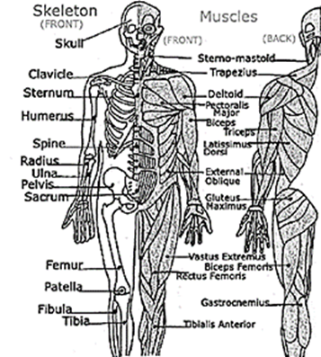
Vertebrates move by the actions of muscles on bones. Tendon attach many skeletal muscles across joints, allowing muscle contraction to move the bones across the joint. Muscles generally work in pairs to produce movement: when one muscle flexes (or contracts) the other relaxes, a process known as antagonism.
Muscles have both electrical and chemical activity. There is an electrical gradient across the muscle cell membrane: the outside is more positive than the inside. Stimulus causes an instantaneous reversal of this polarity, causing the muscle to contract (the mechanical characteristic) producing a twitch or movement.
Skeletal Muscle Structure
- Muscle fibers are multinucleated, with the nuclei located just under the plasma membrane. Most of the cell is occupied by striated, thread-like myofibrils. Within each myofibril there are dense Z lines. A sarcomere (or muscle functional unit) extends from Z line to Z line. Each sarcomere has thick and thin filaments. The thick filaments are made of myosin and occupy the center of each sarcomere. Thin filaments are made of action made anchor to the Z line.
- Muscles contract by shortening each sarcomere. The sliding filament model of muscle contraction has thin filaments on each side of the sarcomere sliding past each other until they meet in the middle. Myosin filaments have club-shaped heads that project towards the actin filaments.
- Myosin heads attach to binding sites on the actin filaments. The myosin heads swivel toward the center of the sarcomere, detach and then reattach to the sarcomere, detach and then reattach to the nearest active site of the actin filament. Each cycles of attachment, swiveling, and detachment shortens the sarcomere 1%. Hundreds of such cycles occur each second during muscle contraction.
- Energy for this comes from ATP, the energy coin of the cell. ATP binds to the cross bridges between myosin heads and actin filaments. The release of energy powers the swiveling of the myosin head. Muscles store little ATP and so must recycle the ADP into ATP rapidly. Creatine phosphate is a muscle storage product involved in the rapid regeneration of ADP into ATP.
- Calcium ions are required for each cycle of myosin-actin interaction. Calcium is released into the sarcomere when a muscle is stimulated to contract. This calcium uncovers the actin binding sites. When the muscle no longer needs to contract, the calcium ions are pumped form the sarcomere and back into storage.
Contraction of Nonmuscular cells
- Action and myosin, whose interaction causes muscle contraction, occur in many other cells. Actin is attached to the inner surface of the plasma membrane. The interaction of cytoplasmic myosin and this actin causes contraction of the cell, such a the coordinated contractions of intestinal cells to absorb nutrients.
- Some fish have modified muscles that discharge electricity. These fish have electric organs consisting of modified muscles known as electroplates. The South American electric eel has more than 6000 plates arranged into 70 columns. Maximum discharge is 100 watts.
Interaction of the Two Systems
- Vertebrates move by applications of the principles of the lever. Levers amplify or increase the force or velocity of motion.
- The amount of amplification depends on the length of the lever. There are three types of skeletal system, all interact with muscles using the lever.