Tissue, Physiology of Plants and Animals
Category : UPSC
TISSUE, PHYSIOLOGY OF PLANTS AND ANIMALS
TISSUE
Tissue is a group of cells with common origin, structure and function that work together to perform a particular function. For example. Blood, bone, cartilage are some examples of animal tissues while xylem, phloem, parenchyma etc. are different types of tissues found in plants. The study of tissue is called histology. The term was coined by Meyer.
PLANT TISSUES
On the basis of their ability to divide, plant tissues are divided into two types:
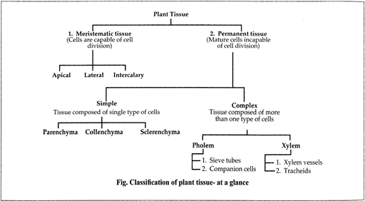
Meristematic Tissues
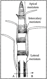
Classification of meristematic tissue
|
On the basis of origin and development |
||
|
Promeristem |
Primary meristem |
Secondary meristem |
|
· Represents primary stages of rneristematic cells. · present at the tip of radicle and plumule |
· Originate from promeristem that continue to divide to form different tissues. · Always in active state of division and give rise to primary permanent tissues for growth in length as well as width e.g., apical meristems, intercalary meristems, lateral meristems (intra- fascicular cambium in the vascular bundle of dicot stem). |
· Developed from primary permanent tissue. They are developed at a later stage by differentiation and acquire power of division, e.g., inter fascicular cambium in stem, cambium in roots and also cork cambium (phellogen). |
|
On the basis of function |
||
|
Protoderm |
Periblem |
Procambium |
|
· It is the outermost layer meant for producing the single layered epidermis, hairs, velamen, stomata i.e., epidermal tissue system. |
· It produces hypodermis, cortex and endodermis or ground tissue system. |
· It is the innermost part of the meristem. It gives rise to the stele which comprises primary vascular tissues and ground tissues like pith, medullary rays and the pericycle. |
PERMANENT TISSUE
Permanent tissue are tissues that have lost the ability to divide, and have attained a definite form and size. They are actually derived from Meristematic cells. Different type of permanent tissues is formed due to differences in their specialization. Differentiation is the process whereby cells take up a definite shape, size, structure and function. These tissues are divided into simple, complex and special.
Table: Difference between Meristematic tissue and Permanent tissue
|
SI. No. |
Meristematic Tissue |
Permanent Tissue |
|
1. |
Meristematic tissues are composed of cells that divide continuously. |
Permanent tissues are composed of cells that are derived from Meristematic tissue. |
|
2. |
Cells are small, undifferentiated and isodiametric in shape. |
Cells are large, differentiated with different shapes. |
|
3. |
Cell wall is thin and living. |
Cell wall may be thin (living) or thick (dead). |
|
4. |
Cells are compactly arranged without inter-cellular spaces. |
Intercellular spaces are often present. |
|
5. |
Nucleus is large and prominent. |
Nucleus is less conspicuous. |
|
6. |
Cells of Meristematic tissue take part in growth. |
Permanent tissue provides protection, support, conduction of substances, storage, photosynthesis etc. |
Simple Tissue
It is made up of only one kind of cells forming a uniform mass. The cells are similar in structure, origin and function. Simple permanent tissues are of three types: Parenchyma, Collenchyma and Sclerenchyma
|
Parenchyma |
Collenchyma |
Sclerenchyma |
|
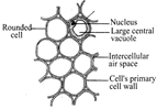
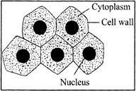
Parenchyma: Parenchyma is widely distributed plant body such as stem, roots, leaves and flower. · They are found in the cortex of root, ground tissue in stems and mesophyll of leaves, cells are isodiametric. · It may contain chlorophyll. Parenchyma containing chlorophyll is called chlorenchyma. It is the site of photosynthesis. · Parenchyma that encloses large air cavities is known as aerenchyma. Aerenchyma provides buoyancy to aquatic plants. |
Collenchyma: Collenchyma is a strong and flexible tissue that provides flexibility to soft aerial parts. · Collenchyma provides mechanical support, flexibility to soft aerial parts so that they can bend without breaking and may contain chloroplasts and thus take part in photosynthesis. |
Sclerenchyma: It is found in and around the vascular tissue, under the skin i.e. the epidermis in dicot stems. · Cells are long, narrow, thick and lignified usually pointed at both ends. The cell wall is evenly thickened with lignin. Lignin is a water proof material. · It gives mechanical support to the plant by giving rigidity, flexibility and elasticity to the plant body.. |
|
|
|
|
Complex Tissue
Complex tissue is made up of more than one type of cells that work together to perform a particular function. Complex tissues are of two types: Xylem and Phloem.
Table: Difference between xylem and Phloem
|
Xylem |
Phloem |
|
Xylem helps in conduction of water and minerals. |
Phloem helps in conduction of food materials and organic solutes. |
|
The flow of material is mostly unidirectional. |
The flow of material is bidirectional. |
|
Xylem consists of tracheids, vessels, xylem parenchyma and xylem fibers. |
Phloem consists of sieve tubes, companion cells, phloem parenchyma and phloem fibers. |
|
Conducting elements of xylem are tracheids and vessels. |
Conducting element of phloem is sieve tubes. |
|
Out of four element of xylem, only xylem parenchyma is living, rest three are dead. |
Out of four elements of phloem, only phloem fibers are dead, rest three are living. |
They are scattered throughout the ground tissue of the plant and contain stored organic matter in the form of starch, rubber, tannins, alkaloids, mucilage enzymes, protein, etc.
TISSUE SYSTEM
In higher plants several tissue work together inform of a unit to perform particular function. These tissue have the same origin such tissue form a system which is called tissue system. On the basis of division of labour, tissue categorised by sachs into three different system-epidermal, grand and vascular tissue system.
Stomata
Vascular Bundles
Secondary Growth
ANIMAL TISSUES
The working of animal body is coordinated by tissues and organs present in their body.
On the basis of structure and function, animal tissues are divided into four types
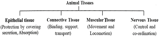
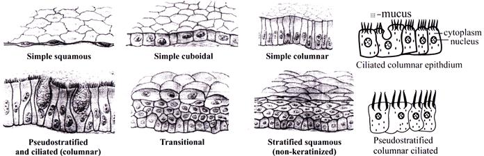
Epithelial Tissue
Epithelial Tissue is the simplest animal tissue that forms the continuous sheet of closely packed cells that covers all external and internal surface of the animal body. Thus, it is also known as covering tissue.
Functions:
|
Compound Epithelium |
|
|
Transitional epithelium |
Stratified epithelium |
|
It is 2-6 cells thick. Basement membrane is absent and occurs in the areas where stretching is required eg. Epithelium of Urinary bladder, Ureter and Pelvis. |
This epithelial layer is more than 6 cells thick Basement membrane is present but single. The name of the epithelium is according to the 'cells' of the top layer. It is of 3-types Stratified Squamous Epithelium is the most common stratified epithelium. The cells of the top layer are squamous. Such epithelium occurs in the region where protection is required, or where there is sufficient wear and tear of the tissue, eg. Buccal cavity (cheek epithelium), skin, pharynx, oesophagus, vaginal epithelium, urethra, conjunctiva and cornea. Stratified Cuboidal Epithelium is present in the ducts of mammary glands and sweat glands. Stratified Columnar Epithelium It mainly occurs in the embryonic tissue and is poorly developed in adults. |
Glandular Epithelium
They are epithelial in origin and therefore, may develop from any of the 3-germ layers.
They can be Exocrine or Endocrine type; Unicellular (ex. Goblet glands) or multicellular type.
Connective Tissue
It is mesodermal in origin. It binds and supports body parts. The cells are loosely arranged, i.e. the intercellular matrix is well developed and Basement membrane is absent. It is nourished with the blood / lymph.
The study of bones is called Osteology and the study of cartilage is called Chondrology.
|
|
BONES |
CARTILAGE |
|
1. |
Outer covering (white fibrous connective tissue) of bones is called periosteum |
Outer covering (white fibrous connective tissue) of cartilage is called perichondrium. |
|
2. |
Bone forming cells are called osteoblasts |
Cartilage forming cells are called chondroblasts. |
|
3. |
Bone Protein is ?Ossein? |
Cartilage protein is ?Chondrin?. (The sugar in cartilage is chondriotin sulphate) |
|
4. |
Osteocytes (bone cells) are solitary |
Chondrocytes (cartilage cells) are in groups of 2?s or 3?s |
|
5. |
Osteocytes are arranged on lamellae |
Chondrocytes are scattered in matrix. |
BLOOD -
Muscular Tissue
Muscular tissue is a contractile tissue that occupies more than 40% of total weight of the body.
Types of Muscle Fibres
On the basis of their location, structure and function, there are three types of muscle fibres.
|
SI. No. |
Striated muscle fibres |
Smooth muscle fibres |
Cardiac muscle fibres |
|
On the basis of structure |
|||
|
1. |
Cells are long and cylindrical in shape. |
Cells are elongated and spindle shaped. |
Cells are small and cylindrical |
|
2. |
Cells are unbranched. |
Cells are unbranched. |
Cells are branched. |
|
3. |
Fibres have blunt ends. |
Fibres have pointed ends. |
Fibres have broad ends. |
|
4. |
Cells are multinucleated. |
Cells are urn-nucleated. |
Cells are uni-nucleated. |
|
On the basis of location |
|||
|
9. |
They are found in limbs, hands, feet, tongue, pharynx etc. |
They are found in urinogenital tracts, digestive tract, lungs, iris, blood vessel etc. |
They are found only in the wall of heart. |
|
On the basis of function |
|||
|
10. |
They are able to perform fast and powerful contractions. Hence, get fatigued soon. |
They perform slow but prolonged contractions. |
They perform powerful and rhyth- mic contraction and get fatigued seldom. |
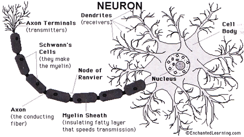
Nervous Tissue
Nervous tissue is specialized to transmit messages in our body. They can receive, integrate and transmit stimuli to various parts of the body. It is devoid of matrix. Its cell is surrounded by a special connective tissue cell. Nervous tissue contains two types of cells Neuron and neuroglial cells.
PHYSIOLOGY OF PLANTS
Morphology is the branch of biology which deals with the study of form, structure and relative position of different organs.
Physiology is the branch of science that deals with the study of different functions of organs.
FLOWERING PLANTS (ANGIOSPERMS)
Herbs: These are the plant, lacking a permanent woody stem and generally dies back at the end of each growing season. E.g., Whean Hen bone, Canna etc.
Shrubs: These are the woody plant which is smaller than a tree and has several main stem arising at or near the ground. E.g., Jasmine rose etc.
Tree: A woody perennial plant having a single usually elongate main stem generally with few or no branches. E.g., Palm, Pinus, Castuarina, Dalbergia, etc.
Creepers: A weak plant that grows along the ground, walls or trees. E.g., grass.
Climbers: They have week stem which help plant to climb up trees and other tall object. E.g., Grape vine etc.,
Lianas: These are the woody climbing plant that hangs from trees, especially in tropical rain forest. E.g., Hiptage, Phanera, etc.
Epiphytes: These are the plant that grow above the ground, supported non parasitically by another plant. E.g., Vanda, etc.
Root System
Characteristics
|
|
Root |
|
||
|
|
\[\downarrow \] |
|
||
|
|
\[\downarrow \] \[\downarrow \] |
|
||
|
Tap root |
Adventitious root |
|||
|
It develops from radicle which is made up of one main branch and other sub-branches. It also forms lateral branches (called secondary roots) which further divide to form tertiary roots. Tap roots, with the secondary and tertiary roots form tap root system. It is the characteristic of dicot plants. |
In some plants after sometime the growth of tap root stops and then roots develops from other part of plant which are branched or unbranched, fibrous or storage, are known as adventitious roots. These are mainly found in monocots and can be grouped into 3 types on the basis of their appearance - roots arising from the base of the stem, e.g. Triticum. - roots arising from leaves, e.g. Bryophyllum. - roots developing from nodes and internodes of the stem.. |
|||
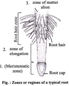
Regions
Modification of roots:
Tap and adventitious roots are modified in different forms to perform special functions and called as modified roots.
|
Modification of Taproot |
Modification of adventitious root |
|
Taproot are modified for food storage and respiration Fusiform roots: Eg. - Radish. Tuberous roots: Eg. Mirabilis. Nodulated roots: Eg. Plants of leguminosae family (Papilionateae) - Pea. Modified tap root for respiration are pneumatophores: The plants, which grow in this region have some branches of tap root that grow vertically upward and comes on surface of soil. These roots are called pneumatophores. They have minute pores called pneumathodes or lenticels by which air enter inside the plant and get oxygen for respiration. Eg. Rhizophora, Mangrove, Heritiera. |
Adventitious roots can be modified on the basis of functions like fleshy for storage (eg. moniliform, annulated, tuberous, fasciculated, palmate nodulose), mechanical support and for vital functions. Tuberous adventitious root: Eg. Sweet potato. Fasciculated roots: Eg. Asparagus, Dahlia. Stilt roots: Eg. Maize, Sugarcane, Pandamis (screwpine). Prop root or pillar roots: Eg. Banyan. Buttress root: Eg. Terminalia. Climbing roots: Eg. Money plant (pothos), Monstera (Betel), Black pepper. Respiratory root: Eg. Avicennia, Jussiaea. Foliar root or Epiphyllous root: Eg. Bryophyllum, Begonia. Sucking or haustorial roots or Parasitic roots: Eg. Dendrophthoe, Cuscuta, Viscum. Annulated roots: Eg. Ipecac. |
Functions of Root:
Economic importance
(i) Sugar beet is edible root, which is an important source of sugar.
(ii) Birth control pills compounds area derived from yam roots.
(iii) Various medicines like ginseng, aconite ipecac, gentian are made from roots
Stem
Characteristics
Functions of stem
Economic importance of stem
(i) Stems like sugarcane are the source of sugar.
(ii) Stems like asparagus, bamboo, etc. are vegetables, cinnamon used as a spice is derived from the bark of the tree.
(iii) Medicines are obtained like quinine, Camphor, etc.
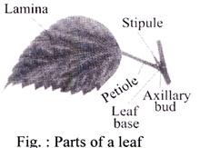
Leaf
|
|
LEAF |
|
|||
|
|
\[\downarrow \] |
|
|||
|
|
\[\downarrow \] \[\downarrow \] |
|
|||
|
Simple |
Compound |
||||
|
It is a leaf which may be incised to any depth, but not down to the midrib or petiole. Eg. mango, guava, papaya etc. |
It is a leaf in which the leaf blade is incised up to the midrib or petiole, thus dividing it into several small parts, known as leaflets. |
||||
Modification of leaves
FLOWER
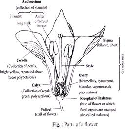
Flower is a specialized branch of limited growth which bears floral leaves that carry on sexual reproduction and give rise to seeds and fruits. The study of flower as called anthology. The part. In a flower, 4 types of floral leaves are found. These are - sepal, petal, stamen and carpel.
|
Flower |
|||
|
Calyx |
Corolla |
Androecium |
Gynoecium |
|
It is the outermost whorl of flower. cach whorl of flower us known as sepal. Sometimes it get modified into spine (Trapa), hairy structure (sunflower), etc. |
It is a second whorl of the flower and each members is known as petals. It may be Polypetlous and gamopetalous. |
It is a male reproductive part of flower. It consists stamen, which is differentiated into anther and filament. |
It is a female reproductive part of flower It comprises stigma, style and ovary. |
RESPIRATION IN PLANTS
Cellular Respiration
Cellular respiration is an enzyme controlled process of biological oxidation of food materials in a living cell, using molecular \[{{O}_{2}},\] producing \[C{{O}_{2}}\] and \[{{H}_{2}}O\] and releasing energy in gradual steps and storing it in biologically useful forms, generally ATP. So respiration is catabolic, exothermic and oxidative process.
\[_{glu\cos e}^{{{C}_{6}}{{H}_{12}}{{O}_{6}}}+_{oxygen}^{6{{O}_{2}}}\xrightarrow[{}]{enzymes}\,_{carbon-dioxide}^{6C{{O}_{2}}}+_{water}^{6{{H}_{2}}O}+_{(ATP)}^{energy}\]
RESPIRATORY QUOTIENT (R.Q.)
The ratio of the volume of \[C{{O}_{2}}\]released to the volume of \[{{O}_{2}}\]taken in respiration is called Respiratory Quotient (R.Q.)
R.Q. = \[\frac{Volume\,\,of\,\,C{{O}_{2}}\,\,evolved}{Volume\,\,of\,\,{{O}_{2}}\,\,absorbed}\]
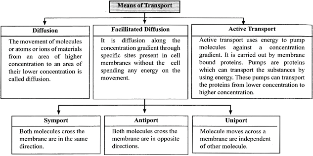
TRANSPORT IN PLANTS
Plants need to move molecules over very long distances, much more than animals do; they also do not have a circulatory system in Place. In a flowering plant the substances that are transported includes water, mineral nutrients, organic nutrients and plant growth regulators.
Plant-water Relations
Water is essential for all physiological activities of plants. It acts as an excellent solvent and help in the uptake and distribution of mineral nutrients and other solutes and also useful for maintaining the turgidity of cells which is essential for cell enlargement, growth & development.
Osmosis
It is a type of diffusion in which water molecules diffuse from the region of higher chemical potential (or concentration) to its region of lower chemical potential (concentration) across a permeable membrane.
Plasmolysis
The behaviour of the plant cells (or tissues) with regard to water movement depends on the surrounding solution. The shrinkage of the protoplast of a living cell from its cell wall due to exosmosis under the influence of a hypertonic solution is called plasmolysis.
|
Solution |
||
|
Isotonic |
Hypotonic |
Hypertonic |
|
If the solution in which a cell is placed, has equal osmotic concentration to that of cell sap, the outer solution is called isotonic solution.
|
If the osmotic concentration of outer solution is less than that of the cell sap, the outer solution is called hypotonic solution. If a cell is placed in such solution endosmosis takes place and cell swells up, e.g., swelling of dried grape (Resins). |
If the osmotic concentration of a solution is Higher than that of the other (cell sap) solution, is known as hypertonic solution.
|
Deplasmolysis:
The swelling up of a plasmolysed protoplast due to endosmosis under the influence of a hypotonic solution or water is called deplasmolysis.
Imbibition
It is a special type of diffusion when water is absorbed by solids - colloids - causing them to enormously increase in volume.
Ascent of sap
Cohesion and tension theory by Dixon and Joly (1894), etc. is the most accepter theory.
Transpiration
Guttation
PLANT GROWTH REGULATORS
Plant hormone is a chemical substance which may be trans-located to another region, for regulating one or more physiological reactions when present in low concentration.
Plant growth inhibitors, e.g., abscisic acid (ABA) and ethylene. They play an important role in plant response to wounds and stresses of biotic and abiotic origin and are involved in growth inhibiting activities such as dormancy and abscission.
|
Plant Disease |
||
|
Diseases |
Causative agent |
Symptoms |
|
Branchy top of Banana |
Banana Virus- 1 |
Newly formed leaves becomes narrow. |
|
Necrosis (potato) |
Potato virus-X(PVX) |
Leaves become dwarf and sharp spots are present |
|
Potato mosaic |
Potato virus-X |
Leaves become dward and exhibits dark spots. |
|
Tobacco Mosaic |
Tobacco Mosaic Virus (TMV) |
Stunted growth of leaves, Mottle appearance and leaves shrinks |
|
Yellow vein Mosaic (Bhindi) |
Begmovirus or (YVM) |
yellowing of veins leaf become chlorotic |
|
· Fungal |
||
|
Early blight |
Alternaria solani |
Concentric rings are present on leaves and it becomes brown, angular, have necrotic spots on the leaves. |
|
Late blight Potato/Tomato |
Phytopthora infestans |
appearance of brown lesions on leaves and stem. |
|
Rust of wheat |
Puccinia graminis |
appearance of red brown rust on stem and leaves. |
|
Loose smut of wheat |
Ustilago tritci |
Infloresence is affected and shows early burst of fruits wall. |
|
Red rot (Sugarcane) |
Colletotrichum falcatum weat, glomerella cingulata |
Let dries and exhibits brown or reddish nodal region |
|
Citrus canker |
Xanthomonas axonopodis pvcitri, Xanthomonas axonopodis PV aurantifolis |
appearance of lesion is leaves, stem and fruit in lemon plant. |
|
Ring disease (witt of potato) |
Pseudomonas solanacearum |
appearance of share brown ring in xylem of rascular system and callapsing of vascular system occurs. |
|
Bacterial leaf spot |
Xanthomonas compestris |
appearance of translucent spots lettcece-large brown to black circular area. |
PHYSIOLOGY IN HUMANS
Physiology is a branch of science which deals with normal Functions takes place throughout the living system. A living body comprises different system which are separate, but inter connected to each other. It includes Digestion, Respiration, Circulation, Neural and chemical control and co-ordination, excretion and their related disorders. It deals with all metabolic and catabolic processes occurs in the living body. The process of conversion of complex food substances to simple absorbable forms is called digestion.
DIGESTIVE SYSTEM IN HUMANS
The digestion in vertebrates occurs in the digestive tract or alimentary canal. The various parts involved in digestion can be broadly divided in two groups -
Alimentary Canal
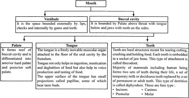
Pharynx
It is a 12 cm funnel shaped passage from buccal cavity to esophagus. It is a common passage for both food and air. A flap, epiglottis closes over the trachea when food is swallowed to prevent choking.
Oesophagus
Stomach
It is differentiated into three main parts, i.e. fundic stomach, body of stomach and pyloric stomach. The fundic as well as body of the stomach are for digestion and contain gastric glands (simple, branched and tubular type).
These glands contain three types of cells.
(i) Mucus cells - They secrete mucus which acts as a lubricant. Mucus also prevents the digestion of stomach by proteolytic enzymes, and injury to stomach, by acid.
(ii) Oxyntic (parietal) cells - They secrete HCl and Castle’s intrinsic factor
(iii) Peptic/zymogen/chief cells -They secrete digestive enzymes.
Functions of HCl-
The secretion of HCl is stimulated by histamine, acetylcholine and gastrin.
Small Intestine
The small intestine is coiled and narrow tube which can be distinctly divided into three regions i.e. Duodenum, Jejunum and IIeum.
(i) Duodenum:
(iii) Jejunum: It is 2.4 metre long and bears finger likes projections called villi which increase the surface area of the inner lining of intestine.
(iii) Ileum: It is 2.4 metre long with club-shaped villi. Its lower end forms a Merkel’s diverticulum. The opening of ileum in caecum (large intestine) called ileocaecal orifice.
Large Intestine
Digestion in Man
Renin enzyme present in infants or curdles milk. Pepsin break proteins into proteoses and peptones.
(i) Trypsin breaks proteins, peptones and peptides into ammo acids.
(ii) Amylase breaks starch into sugar.
(iii) Lipase breaks patsineo acids and glycerol.
NUTRITIONAL AND DIGESTIVE DISORDERS
RESPIRATORY SYSTEM
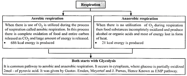
Respiration is an oxidative process occurring within living cells by which the chemical energy of organic molecules is released in a series of metabolic steps involving the consumption of oxygen and liberation of carbon dioxide and water. As the process of respiration takes place inside the cells, it is also known as cellular respiration.
Respiration is of 2-types — aerobic and anaerobic respiration
|
Respiration |
|
|
Aerobic respiration |
Anaerobic respiration |
|
· It is a process of cellular respiration that uses oxygen in order to break down respiratory substrate which then releases energy. · 38 molecules of ATP are released for every glucose molecule broken down. · It takes place in the cytoplasm (glycolysis) and mitochondria (Krebs and Electron Transport Chain) of the cell. · The equation of aerobic breakdown of glucose is: \[_{(6-C\,\,compound)}^{Glu\cos e}\xrightarrow[In\,\,cytoplasm]{Glycolysis}_{(3-C\,\,compound)}^{Pyruvic\,\,Acid}\xrightarrow[In\,\,mitochondria\,\,presence\,\,of\,{{O}_{2}}]{Krebscycle}\] \[C{{O}_{2}}+{{H}_{2}}O+38ATP\] |
· It is a process of cellular respiration that takes place in absence of oxygen, there is incomplete breakdown of respiratory substrate and little energy is released. · 2 molecules of ATP are released for break-down of every glucose molecule. · It takes place in the cytoplasm of the cell, mitochondria is not involved. The equation is: \[_{(6-C\,\,compound)}^{Glu\cos e}\xrightarrow[In\,\,cytoplasm]{Glycolysis}_{(3-C\,\,compound)}^{Pyruvic\,\,Acid}\xrightarrow[In\,\,cytoplasm\,\,of\,\,muscle\,\,cells]{Nooxygen}\] \[Lactic\,\,Acid\] \[(3-C\,\,compound)+2ATP\] |
TYPES OF RESPIRATION AND RESPIRATORY ORGANS OF ANIMALS
Respiratory System in Human
The primary structure involved in respiratory system are lungs. Which are endodermal in origen. Its components are Nasal passage, pharynx, Larynx trachea, Bronchi. Bronchioles and alveoli.
|
Mechanism of Breathing The physical movements associated with the gaseous exchange are called breathing. They are controlled by the respiratory centre of medulla oblongata in the human brain. Thus, the breathing movements are involuntary to a large extent. However, we can control the rate of breathing and the extent of breathing but not for a long time. The respiratory centre is stimulated by the carbon I dioxide concentration of the blood. There are two types of physical movements associated with the gaseous exchange. |
|||||
|
|
\[\downarrow \] |
|
|||
|
|
\[\downarrow \] \[\downarrow \] |
|
|||
|
Inspiration or Inhalation Aspiration of air occurs when the volume of the thoracic cavity is increased. When the volume increases, the pressure in the thoracic cavity becomes lower than the outside atmospheric air. Hence atmospheric pressure forces air into the lungs through the nose and trachea. |
Expiration or Exhalation When the volume of thoracic cavity is reduced, the pressure of the air inside the thoracic cavity becomes greater than outside atmosphere. Hence, air from inside the lungs expelled through the trachea and nose to the outside to equalize the internal and external pressure. |
||||
DISORDERS OF RESPIRATORY SYSTEM
(i) Asbestosis - Exposure to the fibrous minerals of asbestos
(ii) Bauxite fibrosis - Exposure to bauxite fumes that contains aluminium and silica particles.
(iii) Siderosis - due to the deposition of iron in tissue.
(iv) Byssinosis - Also known as “brown lung disease” and caused due to exposure to cotton dust in inadequately ventilated environments.
Bronchitis: It is caused by the permanent swelling in bronchi. As a result of bronchitis cough is caused and thick mucus with pus cells is spitted out. The patient experiences difficulty in breathing.
Tuberculosis (TB): It is caused by bacteria Mycobacterium tuberculosis.
Lung cancer: It is believed that by excess smoking, lung cancer (carcinoma of lungs) is caused. The tissue increases limitlessly, which is called malignancy.
HUMAN CIRCULATORY SYSTEM
Heart
Rhythmicity of Heart
Blood Pressure
The pressure exerted by the blood on the wall of the blood vessels in which it is present is called blood pressure.
It is usually measured in brachial artery by an instrument called sphygmomanometer.
|
|
Artrial blood pressure |
|
||||
|
|
\[\downarrow \] |
|
||||
|
|
\[\downarrow \] \[\downarrow \] |
|
||||
|
Systolic |
Diastolic |
|||||
|
· Systolic blood pressure is the pressure exerted by blood on the walls of the blood vessels due to the systole of ventricles and is equal to 120 mm Hg. |
· Diastolic blood pressure is the pressure exerted on walls of blood vessels when the ventricles are relaxed. During ventricular diastole, the uncoiled elastic layer recoils leading to normalization of artery. Hence, blood pressure drops down to 80 mm Hg. |
|||||
ELECTOR CARDIOGRAM (ECG)
EXCRETORY SYSTEM
Human Excretory System
Excretory system consists of a pair of kidneys, one pair of ureters, a urinary bladder and a urethra.
Kidneys
These are two bean- shaped purplish brown colored structures located in the back of the abdominal cavity. it is the main organ of excretion through which the nitrogenous waste are eliminated in the form of urine about 12 cm long, about 6 cm thick and weight about 150 gm. Kidneys contain millions of nephron which filter 170 to 200 litres blood to produce 1-1.8 litres of urine daily.
Renal Arteries
Two renal arteries constantly transport blood to each of the kidneys.
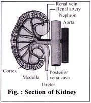
Renal vein
Two renal veins return useful nutrients back into the bloods stream after filtering the unwanted materials in kidneys.
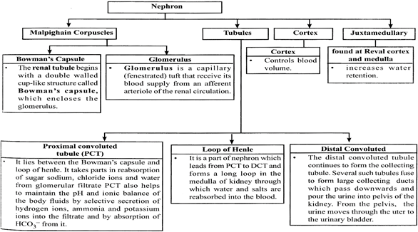
Nephron
There are two types of nephron according to their position in kidney - cortical and juxta medullary nephron.
Dialysis
Urea is a toxic chemical. When it is not removed from the body, due to kidney disease, it gets accumulated in blood (Uremia) and can cause kidney failure. The urea can be removed from the blood by dialysis.
It is less costly but risky as there are chances of infection due to the permanent tube in the abdomen. A fluid (dialysate), containing sodium, chloride, bicarbonate and high percentage of glucose, is introduced into the abdominal cavity through a permanent- attached tube. The peritoneum of abdomen acts as a membrane and the exchange of substances occur with the blood. The fluid, containing urea, is removed periodically.
CONTROL AND COORDINATION IN ANIMALS
DIVISION OF HUMAN NERVOUS SYSTEM
The human nervous system consists of: Central Nervous System (CNS) and Peripheral Nervous System (PNS)
Central Nervous System
It lies in the mid-dorsal region along the lontudinal axis of the body. It consists of two parts. Brain and Spinal Cord.
Brain
This is the highest coordinating centre in the body. It is situated in the head region, in the cranial cavity of the skull. It is soft, whitish organ which weighs 1.2 - 1.4 kg. It forms 98% of the weight of the whole CNS. Brain is surrounded by three protective membranes called. The space between these meninges is filled with cerebrospinal fluid which protects the brain from mechanical shocks. Brain is divisible into three main regions: Fore brain, Mid brain and Hind brain
(i) Fore brain forms the greatest part of the brain. It consists of three regions:
Olfactory lobes are a pair of club-shaped small structures present below the cerebral hemisphere. Both lobes are widely separated. It is centre of smell.
Cerebral hemispheres or cerebrum: It forms the largest part of the brain. It cerebrum has two cerebral hemispheres which lie side by side and are separated by a deep cerebral fissure. The surface of cerebral hemisphere has grooves (sulci) and folds (gyri) to accomodate larger number of nerve cells.
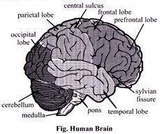
Diencephalon: It is smallest and unpaired part of brain. It lies on the lower side of cerebrum.
(ii) Mid brain: It extends from the pons to the lower portion of the diencephalon. Mid brain is sub divided into Optic Lobes and Crura Cerebri
(iii) Hind brain consists of three parts:
Spinal Cord
It lies in the mid-dorsal region along the longitudinal axis of the body. It is a slender, cylindrical structure, about 45 cm long, originating from medulla oblongata and extending downwards up to the lumber region. Spinal cord is also covered by three meninges, like the brain, in between which is the cerebrospinal fluid. It acts as a centre for reflex actions, thus, reduces brain’s work. It also conducts sensory and motor impulses to and from the brain.
Function:-


You need to login to perform this action.
You will be redirected in
3 sec