Biological Classification/Cell & Its Division
Category : UPSC
BIOLOGICAL CLASSIFICATION/CELL & ITS DIVISION
BIOLOGICAL CLASSIFICATION
Biological classification is the scientific procedure of arranging organisms into groups and subgroups on the basis of their similarities and dissimilarities and placing the groups in a hierarchy of categories. The purpose of biological classification is to organize the vast number of known plants and animals into groups that could be named, remembered and studied.
NEED FOR THE BIOLOGICAL CLASSIFICATION
Classification is needed to
Systematics
Systematics is the study of the units of biodiversity. It is the study of the diversification of organisms and their relationship among living things through time. It includes the following parts:
Classification of Organisms
It is the arrangement of organisms into taxonomic group according to their similarities and dissimilarties. System of classification is an attempt to organize different organisms into different categories that we can use to study.
Hierarchy in Classification
Hierarchy in classification involves many steps. Each step represents a rank or category. All categories or steps together constitute the taxonomic hierarchy.
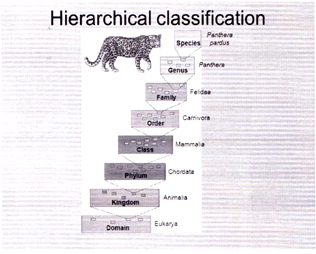
Species
The smallest tax on is species. At the species level organisms look alike and are able to breed with one another.
Genus
The next largest taxon is genus. At the genus level, there is a group of similar species that are closely related.
Family
A group of two or more genera with common characteristics make a family. For example, lion (Panthera leo), tiger (Panthera tigris) and the domestic cat (Felis domesticus) make the family Felidae.
Order
A group of related families make an order. For example the family of cat (Felidae) and the family of dogs, foxes, etc. (Canidae) is grouped under the Carnivora.
Class
Related orders make a class. For example several orders like those of the tigers, cats, dogs, monkey, bats and human belong to class Mammalia.
Phylum
A phylum is the largest category with related classes grouped together. For example the classes of mammals, birds, reptiles, amphibians and fishes together constitute the phylum Chordata. In plants, the corresponding category is named division.
Kingdom
Kingdom is the largest group of organisms differentiated on very general similarities. For example, plant and animal kingdom. The plant kingdom comprises all kinds of plants while animal kingdom comprises all kinds of animals.
Handy Facts
(i) Hierarchical Classification System
(ii) System of Binomial Classification
BINOMIAL SYSTEM OF CLASSIFICATION
Biologists have devised a technique for identification naming and grouping of various organisms.
There is a need to standardize the naming of living organisms such as particular organism. Carl Von Linnaeus devised a binomial system of nomenclature in which an organism is given two names.
FIVE KINGDOM CLASSIFICATION
This type of classification was proposed by R.H. Whittaker. The five kingdom proposed by Whittaker are Monera, Protista, Fungi, Plantae, Animalia.
Kingdom Monera
Economic importance of bacteria
|
Handy Facts |
|
|
Streptomycin |
Streptomyces griseus |
|
Chloramphenicol |
S. venezuelae |
|
Tetracyclines |
S. aureofaciens |
|
Terramycin |
S. ramosus |
|
Erythromycin |
S. erythreus |
|
Bacitracin |
Bacillus Licheniformis Vitamins |
|
Riboflavin |
Clostridium butylicum |
|
Cobalamin (Vit. \[{{B}_{12}}\]) |
Bacillus megatherium |
|
Vitamin C |
Escherichia coli |
Kingdom Protista
Protista are considered as a diverse group of eukaryotic organism. Protists can be unicellular or multicellular and also exists in colonial form. They do not have specialized tissue organization. Protists live in water, in moist terrestrial habitats, and as parasites and other symbionts in the bodies of multicellular eukaroytes.
These are classified into 3 groups :- protistan algal, slime molds, protozoan protists.
|
Handy Facts Protozoan diseases |
|
|
Causative agent |
diseases |
|
· Trypanosome gambiense
|
central african sleeping sickness
|
|
· Giardia Lambia |
Giardiasis |
|
· Leishmania donovani |
Kala-Zar |
|
· Dermal leishmaniasis |
Leishmania tropica |
|
· Plasmodium vivex |
Malaria |
|
· Entamoeba histolytica |
Amoebic dysentery |
|
· Entamoeba gingivalis |
Pyorrhoea |
Economic Importance of Diatoms
Kingdom Fungi
Fungi are basically multi-cellular. Yeast is an exception in being unicellular. The cell wall is generally composed of chitin (a nitrogen containing carbohydrate). They do not contain chlorophyll and hence are heterotrophic. Most of them are decomposers, hence fungi are also known as kingdom of multicellular decomposers. They may be saprophytic (depend on dead or decaying organic matter for their food) or may be parasitic depend on living organisms for their food). Kingdom is classified on the basis of Morphology of reproductive structures, which exhibits more variation.
These are classified into 4 groups. Deuteromycetes, Oomycetes, Zygomycetes, Ascomycetes and Basidiomycetes.
Handy Facts
Lichens are dual organisms that are formed by permanent symbiotic association between an algae and a fungus. They co-exist for mutual benefit. This type of relationship is known as symbiosis. The alga manufactures food for itself and for the fungus. Fungus provides protection to alga and helps in fixation and absorption of water and minerals.
Economic Importance of Lichen
Algal Bryophyta
Red, Green, Liverwort
Brown Hornwort
Handy Facts
Mycorrhizae are the type of symbiotic association between fungus with the root of higher plants, in which both the organism are mutually benefited, these fungus secretes antimicrobial substances, that protects the plant root from harmful pathogens, fungus helps plants to absorb water and important nutrients from the soil. Fungus also derives nutrient from the roots.
Eg. Pinus, birch, etc.
Disease caused by fungi in Humans
|
Allergies
|
Alternaria, phoma, Trichoderma, Aspergillus, etc. |
|
Ear infection |
Aspergillus flavus |
|
Valley fever |
Colcidioidomycosis |
|
Neurites |
Mucor pusillus |
|
Candidiasis |
Candida albicans |
Kingdom Plantae
They are multicellular eukaryotes. All plants contain plastids. Plastids are double membrane organelle that possesses photosynthetic pigments. They are called chloroplast. They are usually autotrophic. Chloroplast contains a green colour pigment called chlorophyll and prepares own food by the process of photosynthesis. Cells have cell wall made up of cellulose.
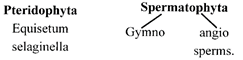
Kingdom plantae shows a lot of diversity, because of which, it has been divided into four divisions. Algae, Bryophyta, Pteridophyta, and Spermatophyta (Gymnosperms and Angiosperms).
These are classified into 4 groups -:
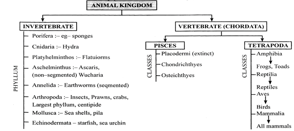
Kingdom Animalia
There are diverse group of animals in the whole world they lives in different habitats. All animals are multicellular except protozoa, these are eukaryotic, lacks cell wall, heterotrophic, have power of locomotion and shows increased sensitivity through the nervous system. On the basis of presence and absence of vertebrate column. Animal Kingdom are broadly divided into vertebrates and invertebrates. Inverbrates consists 8-phylums named Porifera, Cnidaria, Platyhelmintus, Aschelminthus, Anellida, Arthropoda, Molluscs and Enchinodermata. Vertebrates or phylum chordata consists 3-subphylums, Urochordata, Cephalochordata and Vertebrata.
CELL: FUNDAMENTAL UNIT OF LIFE
Cell is a structural and functional unit of life. In 1665, Robert Hooke, an English scientist, saw cells for the first time in a thin slice of cork with its microscope. He observed and described the cells as “Honey comb” like structures. He named the box-like compartments as cellulae or cells. The term “cell” is derived from a Latin word cella which means little room or hollow space.
In 1674, Van Leeuwenhoek, a Dutch Scientist, studied living cells for the first time with the help of an improved microscope.
CELL THEORY
Cells theory, therefore, states that
iii. Cells develop from pre-existing cells. Virus is an exception to cell theory.
|
|
Prokaryotic cell |
Eukaryotic cell |
|
1. |
The size cell is small (0.1-5. \[0\mu m\]). |
Cell Size is larger (5-100\[\mu m\]). |
|
2. |
It consists one envelope organization. |
It consists two envelope organization. |
|
3. |
Unorganized nucleus is present. Hereditary material lies freely in cytoplasm. |
Well-developed organized nucleus is present. Hereditary are embedded in nucleus covered by nuclear membrane. |
|
4. |
Membrane bound organelles like ribosomes, nucleus, endoplasmic-reticulum, golgi body, mitochondria, lysosomes, vacuoles etc. are absent. |
Membrane bound organells are present. |
|
5. |
DNA is naked. |
DNA is associated by histone proteins. |
|
6. |
Site of translation and transcription is cytoplasm ex- Bacteria, Cynabacteria, etc. |
Site of Translation is cytoplasm and Transcription is nucleus ex-plants, animals, fungi. |
|
7 |
Endocytosis and exocytosis does not takes place. |
Endocytosis and exocytosis takes place in protists and animal cell. |
CELL STRUCTURE
Cell Wall
Bacterial cell wall is made up of peptidoglycans. The archean cell wall is made up glycoproteins and polysaccharides. The plant cell wall is mainly composed of cellulose hemi-cellulose glycoproteins, pectins and lignin. Animal cell lacks cell wall.
Plant Cell Wall
Plant cell walls are primarily made up of cellulose which is the most abundant micro molecule on the earth. Plant cell wall consists of three layers, the primary cell wall, secondary cell wall and middle lamella.
The middle lamella:
Primary cell wall (0.1-3\[\mu m\])
Secondary cell wall (3-19\[\mu m\])
Handy Facts
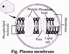
Cell Membrane/Plasma Membrane
The plasma membrane is made up of a bilayer of lipids and proteins. Small carbohydrates are attached at places to outer surface of lipids and proteins.
According to this model:
|
Plant-cell |
Animal-cell |
|
Larger |
Smaller |
|
cell wall present |
Cell wall absent |
|
Plastids present |
Plastids absent |
|
Large one vacuole |
Small vacuoles |
|
Golgi body is present in the form of dictyosomes. |
Golgi body is well developed |
|
Nucleus lies in the peripheral cytoplasm. |
Nucleus lies in center |
|
Centrosome and centrioles are absent |
Centrosome with centrioles are present. |
|
Cannot change shape |
Can change shape |
|
Lysosome absent |
Lysosomes Present |
|
Chloroplast present |
Chloroplast absent |
|
Ribosomes present |
Ribosomes present |
|
ER present |
ER present |
|
Cell wall and plasma membrane both are present |
Only plasma membrane present |
|
Microtubules or microfilaments are Present |
Present |
|
Cytoplasm present |
Present |
Function of cell membrane
CYTOPLASM
It is living portion or protoplasm of cell that comprises gelly like Substance called cytosol and organells with nucleus. It is present in both plant and animal cell. It includes, ER, Golgi bodies, Plastids, lysosomes, peroxisomes, ribosomes, Mitochondria, and Centrosomes.
Endoplasmic Reticulum
Endoplasmic reticulum is a complex network of membrane bound structure which runs through the cytoplasm. Cisternae are spaces within the folds of the ER membranes. It is connected to both the outer nuclear membrane as well as cell membrane. The membrane has the same structure as the plasma membrane but ribosomes do not have membranes.
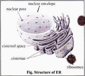
(i) Rough Endoplasmic reticulum (RER): It is lined with ribosomes and is rough in appearance, hence, named as rough endoplasmic reticulum. It is the site of protein synthesis.
(ii) Smooth Endoplasmic reticulum (SER): It contains no ribosomes and hence is smooth in appearance. It helps in lipid and steroid synthesis.
Functions of endoplasmic reticulum:
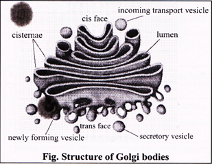
Golgi Bodies
Golgi body consists of smooth, flattened, membrane bound, sac-like structures called cisternae. The cisternae are stacked together; placed one above another in parallel rows. It is Golgi body is a single complex in animal cells while in plant cells, it is formed of separate units called dictyosomes. Membranes of Golgi body may develop connections with membranes of ER to form complex called extra membrane system.
Functions of golgi body
Lysosomes (Lysis = Breaking down; Soma = Body):
Lysosomes are small, spherical vesicle covered by a single membrane. It is scattered all over the cytoplasm. It contains powerful digestive enzymes (about 40 in number) that are capable of breaking down the organic material. Thus, lysosome serves as an intracellular digestive system, and is called digestive bags. These are also known as suicidal bags.
Functions of lysosomes:
Vacuoles
Vacuoles are membrane bound fluid-filled cavities or sacs present in the cytoplasm. They are surrounded by a membrane called tonoplast. The vacuole is filled with a liquid called “cell sap” that contains dissolved salts and sugars.
Functions of vacuoles
Mitochondria
Mitochondria are rod shaped cell organelles surrounded by a double membrane. The outer membrane is smooth and porous while the inner membrane is folded into large number of finger like structures called cristae. Cristae increase the surface area of the inner membrane, which provides more surface area for the metabolic reactions to take place. The fluid inside the mitochondria is called the matrix.
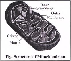
Mitochondria are commonly known as “Powerhouse of the cell”. They contain enzymes necessary for the total oxidation of food and for the release of large amount of energy in the form of ATP molecules. The energy stored in this ATP is used for synthesis of new products and other metabolic process.
Function of Mitochondria
Plastids
(i) Amyloplast: Synthesize and store starch grains.
(ii) Elaioplast (Lipidoplast, Oleoplast): They store lipids and oils.
(iii) Aleuroplast (Proteinoplast): Store proteins.
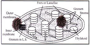
Grana: Inner plastidial membrane of the chloroplast is invaginated to form a series of parallel membranous sheets, called lamellae, which form a number of oval – shaped closed sacs, called thylakoids.
Stroma: It is transparent, protein aceous and watery substance. Dark reaction of photosynthesis occurs in this portions.
Functions of Plastids
Ribosomes
Functions of ribosomes
Cytoskeleton
In eukaryotic cell, a framework of fibrous protein elements became necessary to support the extensive system of membranes. These elements collectively form cytoskeleton of the cell. There are of three types- Microtubules, Microfilaments and intermediate filaments.
Microtubules
The microtubules are electron-microscopic structures found only in the eukaryotic cellular structures like cilia, flagella, centriole, basal-body, astral fibres, spindle fibres. These are mainly formed of tubulin protein.
Functions of microtubules
Microfilaments
These are microscopic, long, narrow, cylindrical, non-contractile and proteins structures found only in the eukaryotic cytoplasm. These are present in the microvilli, muscle fibres (called myofilaments) etc. But these are absent in prokaryotes. These are mainly formed of actin-protein (contractile).
Functions microfilaments
Intermediate Filaments
They are supportive elements in the cytoplasm of the eukaryotic cells. They are missing in mammalian RBCs. The IFs are somewhat larger than the microfilaments and are about 10nm thick. They are solid, unbranched and composed of nonmotile structural proteins, such as keratin, desmin, vimentin.
Functions of intermediate filaments
Functions
CILIA AND FLAGELLA
Centrosome and Centriole
Centrosome is an organelle usually containing two cylindrical structures called centrioles. They are surrounded by amorphous pericentriolar materials. Both the centrioles in a centrosome lie perpendicular to each other in which each has an organisation like the cartwheel. They are made up of nine evenly spaced peripheral fibrils of tubulin.
Functions of centrosome
Nucleus
Nucleus is the prominent, spherical structure found at the center of the cell. It is the largest organelle present in cell. Basically, nucleus is the controlling centre of all cell activities and hence, it has been described as the brain of the cell.
In plant cell, nucleus lies towards the periphery due to the presence of large central vacuole while in animal cell, nucleus lies in the central position.
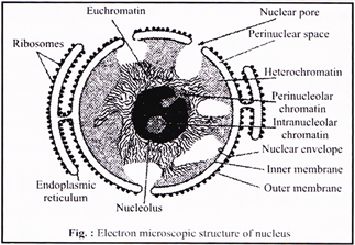
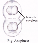
Nuclear envelope or membrane
Nuclear membrane or nuclear envelope, consists of two parallel membranes inner and outer with a space between 10 to 50 nm called the perinuclear space, forms a barrier between the materials present inside the nucleus and that of the cytoplasm. The outer membrane usually remains continuous with the endoplasmic reticulum and also bears ribosomes on it.
Functions:
The nucleolus
Nucleolus is a conspicuous, darkly stained spherical body found in nucleoplasm. It is composed of large amount of ribosomal proteins and ribosomal RNA. It is generally associated with nucleolar organizer region (NOR) of the nucleolar chromosomes.
Functions of nucleolus
Nucleoplasm:
Nuclear matrix:
Function of nuclear matrix
Chromosomes
Functions of Chromatin
Handy Facts
Giant Chromosome
It was discovered by E.G. Balbiani in 1881. These are Commonly present in salivary glands of insect, hence known as salivary chromosomes. Its length is 2000\[\mu m\]
Lamp brush chromosome
It was discovered by Flemming in 1882. It is larger in compared to giant chromosome. These are visible in diplotene stage of most animal 006ytes, spermatocytes. And giant nucleus of unicellular algae i.e. Acetabularia.
Micro bodies
Peroxisomes (Uricosomes)
Glyoxysomes
CELL DIVISION
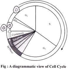
CEIL CYCLE
Phases of Cell Cycle
The period required to complete one cell cycle (from beginning of one cell division to the beginning of next) is called generation nine. It is 24 hours in human cells and 90 minutes in yeast. Cell cycle is simpler in prokaryotes and more complex in eukaryotes.
The cell cycle is divided into two basic phases:
Interphase
Interphase is Completed into Three Successive Stages
Fallowing events take place during this phase
(i) Intensive cellular synthesis.
(ii) Synthesis of rRNA, mRNA ribosomes and proteins.
(iii) Metabolic rate is high.
(iv) Cell size increases.
(v) Synthesis of enzymes, amino acids, nucleotides etc. but there is no change in DNA amount.
S or synthesis phase marks the period during which DNA synthesis or replication takes place.
Following events take place during this phase
(i) DNA replicates and its amount becomes double. If the initial amount of DNA is denoted as 2C then it increases to 4C.
(ii) Synthesis of histone proteins and NHC (non-histone chromosomal proteins).
(iii) Duplication of centriole in the cytoplasm.
G2-phase/Pre mitotic/Post synthetic phase/Gap-II
Following events take place during this phase
(i) Mitotic spindle protein (tubulin) synthesis begins,
(ii) Chromosome condensation factor appears.
(iii) Synthesis of 3 types of RNA, NHC proteins, and ATP molecule.
(iv) Repair of damaged DNA occurs.
|
|
Cell division |
|
||||
|
|
\[\downarrow \] |
|
||||
|
|
|
|
||||
|
|
\[\downarrow \] |
\[\downarrow \] |
\[\downarrow \] |
|
||
|
Amitosis |
Mitosis |
Meiosis |
||||
|
· Amitosis is also called as direct cell division. |
· Mitosis is also called indirect cell division or somatic cell division or equation division.
|
· It is a division that occurs in a mature diploid reproductive cell (2x) in which nucleus divides twice but chromosome (DNA) replicates only once to form four haploid cells, each having half the number of chromosomes present in the parent cell. As it causes reduction in the number of chromosomes, it is known as reduction division.
|
||||
|
· In this division there is no differentiation of chromosomes and spindle. The nuclear envelope does not degenerate. The nucleus elongates and constricts in the middle to form two daughter nuclei. This is followed by a centripetal constriction of the cytoplasm to form two daughter cells. Examples: Prokaryotes, protozoans, yeasts, foetal membrane of mammals, cartilage of mammals etc. |
· In this, mature somatic cell divides in such a way that chromosomes number is kept constant in daughter cells equal to those in parent cell.
|
|
||||
|
|
· The growing regions of plants have meristematic cells (e.g. these cells are found in apical portion of root and stem and in the expanding leaf) in which mitosis takes place.
|
|
||||
MITOSIS
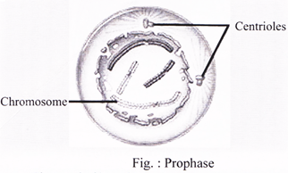
Prophase
It is the longest phase of karyokinesis.
Prometaphase
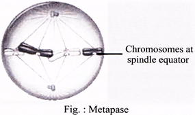
Metaphase
Anaphase

Telophase
Cytokinesis
ENDOMITOSIS
|
S. No. |
Animal cell cytokinesis |
Plant cell cytokinesis |
|
1. |
Centrioles present at spindle poles. |
Centrioles lacking at spindle poles. |
|
2. |
Asters are formed (amphiastral). |
No asters are formed (anastral). |
|
3. |
Cytokinesis by furrowing of cytoplasm. |
Cytokinesis mostly by cell plate formation. |
|
4. |
Furrow extends centripetally. |
Cell plate grows centrifugally. |
|
5. |
Occurs nearly in all tissues. |
Occurs mainly at meristems. |
|
6. |
Cell becomes rounded and its cytoplasm more viscous at the time of mitosis. |
Cell does not change from or nature at the time of mitosis. |
MEIOSIS
|
Meiosis I |
Meiosis II |
|
Prophase I |
Prophase II |
|
Metaphase I |
Metaphase II |
|
Anaphase I |
Anaphase II |
|
Telophase I |
Telophase II |
Meiosis I
It results in the formation of two haploid cells from one diploid cell. The daughter cells are, therefore, haploid but with 2n DNA content. It is divided into four phases i.e., prophase, metaphase, anaphase, telophase.
Prophase-I: It is of longest phase of karyokinesis of meiosis. It is again divisible into five subphases i.e., leptotene, zygotene, pachytene, diplotene and diakinesis.
(i) Leptotene/Leptonema
Chromosomes are long thread like with chromomeres (i.e. linear series of darkly stained swollen areas) on it. homologous chromosomes derived from different parents either paternal or maternal.
(ii) Zygotene/Zygonema
Pairing or “Synapsis” of homologous chromosomes takes place in this stage.
Paired chromosomes are called bivalents, which by further molecular packing and spiralization becomes shorter and thicker.
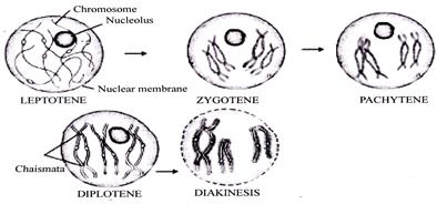
(iii) Pachytene/Pachynema
Crossing over It takes place by breakage and reunion of chromatid segments. Breakage called nicking, is assisted by an enzyme endonuclease and reunion termed annealing is added by an enzyme ligase.
(iv) Diplotene/Diplonema
This stage the paired chromosomes begin to separate (desynapsis) terminalisation starts is formed at the place of crossing over between non-sister chromatids.
Homologous chromosomes move apart they remain attached to one another at specific points called chiasmata.
(v) Diakinesis
Terminalisation of chiasmata. Occurs Nuclear membrane and nucleolus degenerates. Chromosome recondense and tetrad moves to the metaphase plate. Formation of spindle. When the diakinesis of prophase-I is completed than cell enters into the metaphase-I.
Metaphase-I
Chromosomes allign on the equator. Bivalents arrange themselves in two parallel equatorial or metaphasic plates.
Anaphase-I
In involves separation of homologous chromosomes which start moving towards opposite poles so each tetrad is divided into two daughter dyads. So anaphase-I involves the reduction of chromosome number, this is called disjunction.
Telophase-I
Two daughter nuclei are formed but the chromosome number is half of the chromosome number of mother cell. Nuclear membrane reappears and after telophase I cytokinesis may or may not occur.
Significance of meiosis-I
Meiosis-II
It is also called equation or homo typical division because the number of chromosomes remains same as after meiosis-I. It involves the separation of two chromatids of each chromosome and their movement to separate cells. It is divided in four phases i.e., Prophase-II, Metaphase-II. Anaphase-II and Telophase-II.
Significance of Meiosis-II


You need to login to perform this action.
You will be redirected in
3 sec

