Tissues
Category : 9th Class
Tissues
Chapter Overview
You have studied in the previous chapter that all living organisms are made up of cells.
They are either unicellular (e.g. diatoms, bacteria, yeast protozoans etc.), or multicellular (e.g.) frog, earthworm, dog, man, mango tree, money plant, peepal etc). Most of cells are specialized to carry out different functions. Each specialised function is taken up by a different groups of cells. Since these cells carry out only a particular function.
For instance in human beings, muscle cells combine together to perform contraction and relaxation to cause movements, nerve cells cord in ate to carry messages; blood cells and plasma to transport oxygen, carbon dioxide, food, hormones and waste materials, and so on. Similarly in plants, cells combine to perform specific functions such as transportation of food and water from one part to the other; synthesis of food material, storage of reserve foods, etc. Thus a kind of division of labour exists in the cells of multicellular organisms to perform specific functions.
The body of multicellular organisms is made up of organ systems, organ systems are made up of organs, organs are composed of tissues and tissues are composed of cells. Most of these cells are specialised to carry out only few functions efficiently. These functions are taken up by different groups of cells. Thus, we can say that there is a division of labour in the multicellular organisms.
"Division of labour refers to the distribution of different functions among different parts of the body of organism which get specialized for the particular function."
Cell division and cell differentiation lead to the development of specific organs, consisting of specific groups of cells to perform specific functions in the body. Moreover, the organs are also made up of different groups of cells on the basis of their functions. A particular function, inside an organ is performed by group of specialised cells which lie at a definite site in the body. The cluster of cells specially positioned and designed to perform a particular function efficiently is known as tissue.
|
Tissue: A group of similar or dissimilar cells that perform a common function and have a common origin, is known as Tissue |
Cell
\[\downarrow \]
Tissue
\[\downarrow \]
Organ
\[\downarrow \]
Organ System
\[\downarrow \]
Organism
Do You Know
The plants and the animals both have similar life processes. However, they do not have similar types of tissues because of the dissimilarities in their organisation, mode of living and life styles.
Plant on the other hand are autotrophic and, therefore, need a different type of organisation
On the other hand animals are mobile (except sponges and some coelenterates). They dove around in search of food, mate and shelter. Most of their tissues are living. The living tissues require more energy for their maintenance.
Cells of plants exhibit great variation in size and structure. A group of cells, which are all alike in structure and function and have a common origin is called a tissue. In living beings each tissue has its specific function. With the help of different tissues vital activities are performed.
On the basis of development, plant tissues are basically classified into two main groups:
II. Permanent tissue
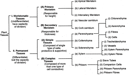
The group or cluster of cells (meristematic cells) which has a capacity of continuous cell division is known as meristematic tissue. Such type of tissues are found in growing regions where cells are divided continuously to attain growth. These cells help in increasing the length and the girth of the plant.
Definition: A group or cluster of living cells which are located at specific locations and divide continuously nucleus to add new cells to plant body is called meristematic.

Fig. 5.1 Meristematic tissue as seen in cross section
Characteristics of the cells of Merestematic tissues
Occurrence: Meristematic tissues are growth tissues and are found in the growing regions of the plant such at the Apies of root and shoot. Depending upon the occurence and position in the plant body, meristems are of three types:
1. Apical Meristem- Such type of tissues are found on apical portions of stems and roots. Due to the continuous divisions of these cells, plant grows in length. About physiology of meristematic tissues many botanists have propounded different theories. Some of them are following:
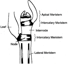
Fig. 5.2: Schematic representation of longitudinal section of stem tip showing location of meristematic tissue in plant body.
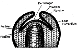
Fig. 5.3: Diagrammatic longitudinal section through shoot apex.

Fig. 5.4: Median longitudinal section of root apex.
3. Intercalary Meristem- in fact, it is also a part of apical meristem which gets separated during root and stem elongation from apical meristems. Such type of meristem is found in internodes of stems and below the nodes of dicotyledonous stems. They are responsible for growth of leaves and internodes.
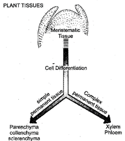
Fig. 5.5:
Permanent tissues are those tissues in which growth has been ceased, at least temporarily.
Permanent tissues as a whole in part may again become meristematic. These are composed of cells which have lost the power of division, having attained their definite form, size and shape.
They may be primary or secondary. The process of taking up a definite shape, size, structure and function is called differentiation.
Definition: The tissues which consists of cells that have lost the ability of dividing and have attained their definite form and size are called permanent tissues.
Characteristics of permanent tissues:
(i) The cells of permanent tissue may be thin or thick walled.
(ii) The cells have undergone differentiation and assumed definite size and function.
(iii) Nucleus is small in relation to cell size and cytoplasm is peripheral with a central vacuole.
(iv) The cells of permanent tissues normally do not divide.
(v) Permanent cells perform specific function.
(vi) The cells may be living or dead.
Classification of permanent tissue:
The permanent tissues are classified into three groups-
Simple Dermanent Tissues
Simple permanent tissues have cells of similar shape, size and structure. They perform a definite function. They have living or dead, thin or thick walled cells. Simple permanent tissues are of following kinds:
General Characters: Parenchyma is most simple and unspecialized primitive tissue mainly consists of thin-walled cells which have intercellular space between them. Parenchyma forms the bulk of the plant body that lies in between specialized tissues. The cells of parenchyma are living. The cell wall is made up of cellulose. The cells have prominent nucleus and large central vacuole in the cytoplasm and perform metabolic processes.
Shape of cells: The cells of parenchyma tissue may be oval, rounded or polygonal, spherical, cylindrical, rectangular, stellate or long spindle like in outline. The cells are isodiametric.
Distribution: The parenchyma is widely distributed in plant organs such as root, stem, leaves, flowers and fruits. The parenchyma is found in soft parts of the plant such as in epidermis, cortex, pericycle, pith, leaf mesophyll, pulp of fruits, and endosperm of seed.
Types of Parenchyma: There are many types of parenchyma. It is precursor of other tissues. When parenchyma tissue contains chloroplasts, it is called chlorenchyma e.g. leaves etc. and is responsible for photosynthesis. Fairly large air cavities may be present in the parenchyma cells of aquatic plants, which provide buoyancy to the plant, such tissue is called aerenchyma.
Idioblasts are specialized parenchymatous cells which produce and store tannins, oils and calcium oxalate crystals.
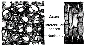
Fig. 6.1.: Parenchyma
Functions:
General Characters: The cells of collenchyma are living and isodiametric or some what elongated. The cells are thin-walled but possess thickenings of cellulose and pectic substances at the corners where number of cells join together. Intercellular spaces are very less or fully absent between the cells. The tissue provides flexibility to soft aerial parts of plant so that they can bend without breaking.
Shape of cells: The collenchymatous cells are generally elongated with oblique end walls.
In transverse section, cells appear, polygonal, circular or oval. Each cell possesses a large central vacuole and a peripheral cytoplasm with a prominent nucleus.

Fig. 6.1: Collenchyma
Distribution: The collenchyma cells are located below the epidermis (i.e. hypodermis) of dicotyledonous stems and leaf petiole (in outer region of cortex). These cells are also found in the mid ribs of dicot leaves. Collenchyma is generally absent in monocot roots, stems, and leaves.
Functions:
Table. 6.2.: Differences between Parenchyma and Collenchyma
|
|
Parenchyma |
|
Collenchyma |
|
1. |
It has isodiametric cells. |
1. |
It has elongated cells. |
|
2. |
The cell wall of parenchyma is uniformly thin. |
2. |
The cell wall get unevenly thickened at comers. |
|
3. |
It is distributed in almost all the parts of the plant body. |
3. |
It occur mostly in the aerial parts of the plants, restricted to the outer layers. |
|
4. |
The living cells of parenchyma assimilate and store food. They also store waste materials. |
4. |
Collenchyma is the chief mechanical tissue in parts of a young plant particularly in the young dicotyledonous stems. |
General Characters: Sclerenchyma is also a simple permanent tissue. The cells are thick walled with little or no protoplasm. Cells are dead at maturity. Sclerenchymatous cells have hard and extremely thick secondary walls due to uniform deposition oflig11111. Lignin deposition is so thick that the cell walls become strong, rigid and impermeable to water. Sometimes oblique thin areas are found in thick cell walls which are called pits. The cells are closely packed without intercellular space in Sclerenchyma. The cells are cemented with the help of a conspicuous middle lamella. The middle lamella is a wall that lies between the adjacent cells. It is made of pactate of calcium and magnessium,
Shape: The cells of Sclerenchyma appears as hexagonal net in transverse section.
Do You Know
Lignin: It is a complex polymeric molecule composed of phenyl propanoid units associated with cellulose. It acts as a cement and hardens cell wall. It provides flexibility and great tensile and compressional strength. A high tensil strength means that it does not break easily on stretching, and a high compressional strength means that it does not buckle easily.
(i) Fibres: Fibres are highly elongated (1-90 cm), narrow and spindle shaped with tapering end walls. The fibres are usually clustered into strands and look polygonal in transverse section.
Adjacent fibres possess simple oblique pits. They are empty and dead at maturity and provide mechanical strength to the base in which they occur.
(ii) Sclereids: These are highly thickened dead sclerenchymatous cells with very narrow cavities. These may occur singly or in groups. They provide stiffness to the plant part in which they occur. They are short and vary greatly in their shape and size. They may be cylindrical, spherical, ovel, T-shaped, tumble shaped or stellate.
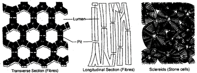
Fig. 6.3: Sclerenchyma
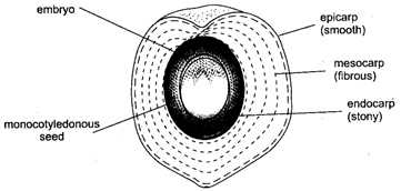
Fig. 6.4. Parenchyma
Distribution: Sclerenchymatous fibres abundantly occur in hypodermis, pericycle, secondary xylem and secondary phloem. They usually occur in patches or in definite layers. On the other hand, the sclereids occur singly scattered in cortex, pith. Phloem etc. They also occur in hard seed coats, hard endocarp of almond and coconut, grit of, pear and sapota.
Functions:
(1) Sclerenchyma saves the plant from various stresses of environmental forces like strong wind etc.
(2) Sclerenchyma in leaves provides rigidity and saves it from collapsing.
(3) Fibers are mainly responsible for providing the plant, mechanical strength. Thus they holy in retaining their position.
(4) Sclerosis provides grittiness in the pulp of many fruits.
Table. 6.5: Differences between collenchyma and sclerenchyma
|
|
Collenchyma |
|
Sclerenchyma |
|
1. |
Collenchyma consists of living cells. |
1. |
Sclerenchyma consists of dead cells. |
|
2. |
Its cells have soft primary walls. |
2. |
Its cells have hard secondary walls. |
|
3. |
Cell walls are thin but thickened at corners due to deposition of cellulose and pectic substances. |
3. |
Cell walls are thick as they have lignin deposition. |
|
4. |
It has same type of cells. |
4. |
It has fibres and sclereids. |
|
5. |
Cells are filled with protoplasm. |
5. |
Cells are empty and have narrow lumen. |
|
6. |
They perform vital functions only |
6. |
They provide strength and rigidity to |
Table 6.6: Differences between fibres and sclereids
|
|
Fibres |
|
Sclereids |
|
1. |
Fibres ate elongated |
1. |
Sclereids are broad. |
|
2. |
They have tapering end walls. |
2. |
The end walls are blunt. |
|
3. |
They are generally unbranched. |
3. |
They are branched or unbranched. |
|
4. |
The pits of fibres are narrow and unbranched. |
4. |
The pits are deep and branched. |
|
5. |
They originate from meristematic cells, |
5. |
They are formed by the thickening of parenchyma cells. |
Protective Tissue
Some cells cover plant parts such as root, stem, leaves, flowers, fruits etc. and provide protection against pathogens and adverse environmental factors. Hence these cells form a protective tissue. Protective tissue includes epidermis and cork.
The epidermis of leaves and green stems is interrupted by small pores, called stomata (sing. stoma; Gk. Stoma = mouth). A typical stoma is microscopic structure and usually consists of two kidney shaped guard cells surrounding an elliptical pore. The guard cells are generally much smaller in size as compared to epidermal cells. The guard cells generally have thick walls towards pore and thin walls towards outer side. This property helps in the opening and closing of stomata. The guard cells have a dense protoplasm, having chloroplasts, a nucleus, mitochondria etc. The stoma helps in transpiration and exchange of gases. In xerophytes stomata are sunken and reduce water loss.
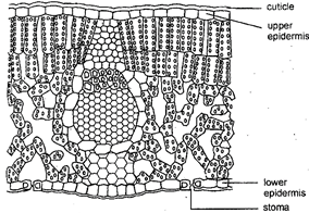
Fig. 6.7: A dorsiventral leaf in transverse section
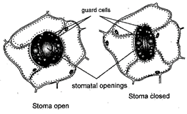
Fig. 6.8: Stomata
The epidermis of leaves and stem may possess unicellular or multicellular hairs or trichomes.
The seeds of cotton possess numerous, long unicellular hairs. The epidermis of young roots possess long unicellular root hairs. They increase the absorption surface and absorb water from the soil. The epidermis in young roots is known as epiblema.
Functions of Epidermis
(i) It gives mechanical support and strength to plant parts.
(ii) It protects the internal tissues from injuries, chemicals and invasion by pathogens.
(iii) It reduces the rate of transpiration by developing cuticle.
(iv) Exchange of gases and transpiration through stomata on leaves etc.
(v) It bears unicellular or multicellular appendages in the form of root hairs, stem hairs, glands etc.
(vi) The hygroscopic velamen and absorbing tissue found in epiphytic roots of some orchids is a part of multilayered epidermis.
2. Cork: It is outer protective tissue of older roots and stems. Cork is originated by a secondary lateral meristem called cork cambium or phellogen. Cork cambium forms secondary cortex i.e. phelloderm on the inner side and cork i.e. phellem on the outer side. The cork cells are dead and do not have any intercellular spaces. They are compactly arranged with their cell walls coated with waxy substance, suberin. The cells are filled with air, tannins and resins. At some places cork possesses small aerating pores called lenticels. Commercial cork is obtained from the stem surface of cork oak (Quercus suber).
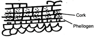
Fig. 6.9: Transverse or cross section of a cork piece showing dead cells
Function
(i) Cork checks the entry of harmful microorganism into plant parts.
(ii) Multilayered impervious cork prevents loss of water by evaporation.
(iii) Cork provides protection against mechanical injury, extreme temperature, fire and browsing animals.
(iv) Cork bears lenticels for exchange of gases between the atmosphere and underlying tissues.
Complex Permanent Tissues
The conducting region of a plant is made up of xylem and phloem. They constitute complex permanent tissue because they have many kinds of cells. In contrast, simple tissues discussed earlier are made up of single type of cells. Common permanent tissue xylem and phloem mainly function as conducting tissues and are known as vascular tissue-
Definition: A complex permanent tissue may be defined as a group of more than one type of cells having a common origin and working together as a unit to perform a common function
The term xylem was introduced by Nageli (1858). Xylem is the chief water conducting tissue of a vascular plant and it also provides mechanical strength to the plant body. Secondary xylem is also called wood. It consists of four types of cells:
(i) Tracheids
(ii) Vessels (or trachea)
(iii) Xylem parenchyma
(iv) Xylem fibres
(i) Tracheids: They are elongated, tubular dead cells. They consists of narrow lumen and moderately thick lignified walls. Thickening are present in various forms such as annular spiral, reticulate, scalar form and pitted. In cross section the tracheids appear angular or polygonal in outline. Tracheids are connected to one another laterally at their ends. Water and minerals are conducted in upward direction from one tracheid to another.
(ii) Vessels: Vessels are much elongated tubes formed of rows of cells called vessel element placed one above the other. They are interconnected through their perforations. Walls of vessels are lignified and are moderately thick. Vessels possess a wide lumen. End walls of the vessels may be oblique or transverse and have open perforated plate i.e. walls of pits completely break down or dissolve. Like tracheids, the thickening in vessels may be annular, spiral, scalarifonn reticulate or pitted. At maturity, nucleus is absent in vessels. Presence of vessels is a characteristic feature of angiospems.
Do You Know
Vessels are present in almost all angiosperms, but there are 10 woody genera in which vessels are absent.
Vessels are usually absent in pteridophytes and gymnosperms but they are found in some species of Selaginella and Pteridium (pteridophyta) and order gnetales of gymnosperms.
(iii) Xylem Fibres: They are the sclerenchymatous cells associated with xylem tissue.
They are more abundant in secondary xylem and provide more mechanical strength. They have extremely thick walls and bear narrow lumen. They are dead cells.
(iv) Xylem Parenchyma (wood parenchyma). It consists of living parenchyma to us cells associated with xylem. The walls are thin and made up of cellulose. The cells are living and serve the function of storage of food. It also helps in lateral conduction of water and minerals.
Wood parenchyma cells are generally elongated in vertical direction in secondary xylem.
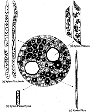
Fig. 6.10: Component cells of xylem tissue
Function of Xylem
Table: 6.11: Differences between Fibers and Tracheids
|
|
Fibres |
|
Tracheids |
|
1. |
They are elongated and tapering sclerenchymatous cells. |
1. |
They are also elongated but shorter than fibres with tapering end walls. |
|
2. |
Fibre cells have a narrow lumen and uniformly thickened cell walls. |
2. |
Tracheidial cells have broad lumen with lignified cell walls showing various types of thickenings. |
|
3. |
They only provide mechanical support. |
3. |
They provide mechanical support and also help in the conduction of water. |
Phloem is a complex permanent tissue responsible for the conduction of organic food materials in the plant body from the region where it is synthesized (source) to the region where it is required (sink). Phloem is called living conducting tissue as it has transport channels made of living cells. It is also called best. Phloem is made up of four types of elements-
(i) Sieve tubes
(ii) Companion cells
(iii) Phloem parenchyma
(iv) Phloem fibres
(i) Seive Tubes: Sieve tube are the long tubular conducting channels which are formed of several cells called sieve tube elements. These cells are placed end to end with their end walls being highly specialized sieve areas called as sieve plates. The sieve plates connects the protoplasts of adjacent seive tube cell. A seive tube cell has peripheral layer of cytoplasm without any nucleus. Nucleus, however, is present in young cells. Each sieve tube cell is accompanied by one or more companion cells. As in mature sieve tube element lacks nucleus it's functions are controlled by a nucleus of companion cell. Sieve pores ir winter get plugged with a substance called callose and hence the transport of food is retarted.
In pteridophytes and gymnosperms, sieve tube elements are not arranged in linear rows, hence are referred to as seive cells.
(ii) Companion cells: These are specialised thin walled parenchymatous cells found associated with sieve tubes. These cells contain dense cytoplasm and prominent nuclei. They are found on sides of sieve tubes. Both sieve tube and companion cells appear from the same mother cell. A companion cell possesses all the important cellular contents such as nucleus (absent in gymnosperms), mitochondria, endoplasmic reticulum etc. The common wall between sieve tube and companion cell shows presence of fine pits which are traversed by Plasmodesmata.
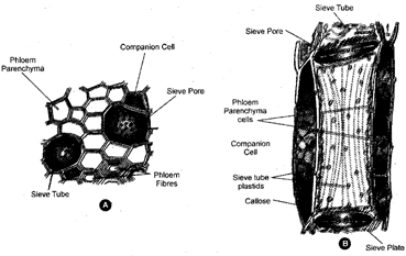
Fig. 6.12: A. Cells of phloem tissue (in T.S.) B. Longitudinal section of phloem showing sieve tube and companion cell
(iii) Phloem Parenchyma: These are thin walled living parenchymatous cells. They are absent in most of monocotyledons and herbaceous dicotyledons. The cells contain cytoplasm and nucleus. They store food materials and may show the presence of crystals, tannins, mucilage, latex etc.
(iv) Phloem Fibres: In primary phloem they are absent or very rare. However they are abundantly present in secondary phloem. They are made up of thick walled sclerenchymatous cells and are mechanical in function. They are also called bast fibres-
Obliteration: After the loss of protoplast, a sieve element is crushed by the pressure of adjoining cells and its contents are absorbed by the adjoining cells. This crushing and absorbing process of sieve elements and companion cells is called obliteration-
Functions of Phloem:
Phloem or best fibres of some plants are source of commercial fibres e.g. flax, jute, hemp etc.
Like plants, the body of multicellular animals consists of different types of tissues which performs specific functions. To understand animal tissues let us take an example. When we breath, our chest moves up and down. This movement is brought about by muscle cells in the body. Continuous contraction and relaxation of muscle cells bring about this movement. When we inspirations we take oxygen. This oxygen is absorbed in alveoli of lungs and is carried to all body parts through blood. In this process various type of muscles and blood take part. In this example both muscles and blood, are example of tissue.
Depending on the basis of their structure and function, animal tissues are of four types-
Characteristics: Epithelial tissue or epithelium is a simplest type of animal tissue. It consists of one or more layers of tightly packed sheets of cells which covers the external surface of the body and viscera (internal organs) and also lines all the body cavities and hollow organs.
For example the skin, of mouth, alimentary canal, lungs etc. The cells are compactly arranged and there is no intercellular space and matrix between them. Cells are held together by intercellular functional complexes. Cells of lowermost layer always rest on a non-living basement membrane or basal lamina which separates it from underlying connective tissue. The epithelium is not traversed by blood vessels. The blood vessels lie in the connective tissue across the basement membrane. Cells have the power of division and regeneration throughout life. Free surface of the cells may be smooth, or may have fine hair-like cilia or microvilli.
Funetions
Types of Epithelial Tissue
On the basis of their structure and functions, epithelial tissues are divided into five groups:
Structure: Squamous epithelium is also known as pavement epithelium because its cells fit together like the tiles of a floor. It is formed of thin, flat, dissocial and polygonal cells with central bulging having flat nuclei. The margins of cells may be smooth or wavy. Squamous epithelium is called simple epithelium when the thin and flat cells form a delicate lining. e.g., lining of capillaries, alveoli etc. Squamous epithelium is called stratified squamous epithelium or multilayered epithelium when cells of skin are arranged in many layers to prevent wear and tear.

Fig. 8.1: Squamous epithelium. A. Surface view; B. Vertical section
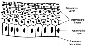
Fig. 8.2: Stratified squamous epithelium
Location: Simple squamous epithelium lines the coelom, blood vessels, urinary tubules, and alveoli of the lungs. Stratified squamous epithelium forms epidermis of the skin. In skin the upper layers of squamous epithelium are keratinesed i.e., have deposition of protein keratin, hence, are dead. In mouth carity the stratified squamous epithelium in non-keratinised. It also lines the buccal cavity, oesophagous, anal canal, vagina and lower part of urethra.
Function: The main function of squamous epithelium is provide protection to the underlying parts against abrasion and entry of germs or chemicals. It also helps in gaseous exchange, ultrafiltration in Bowman's capsule, secretion of coelomic fluid and excretion.
2. Cuboidal Epithelium
Structure: Cuboidal epithelium is formed of compactly arranged cells which appear cuboidal in vertical section and polygonal in surface view. Nucleus of each cell is rounded and centric.
Microvilli may occur on the free surface for increasing absorptive area. Cuboidal epithelium is of two types, simple and stratified.
Location: The cuboidal epithelium occurs in kidney tubules, thyroid vesicles and in glands eg., salivary glands, and exocrine pancreas. It forms germinal epithelium of gonads (testes and ovaries)
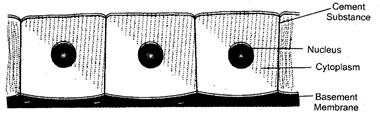
Fig. 8.3: Cuboidal epithelium
Function: Being a surface layer, cuboidal epithelium provides protection to underlying tissues. It also takes part in secretion, excretion and absorption. The germinal epithelium of gonads forms gametes.
Structure: Columnar epithelium is formed of tall, pillar-like cells lying side by side and are polygonal in surface view. Nucleus is longitudinally oval with variable position but generally lies towards the base. Free surface of cells may bear a number of tiny finger like projections called microvilli. Like squamous and cuboidal epithelia, columnar epithelium may be simple or stratified.
Location: Simple columnar epithelium is found in the lining of stomach, intestine and their glands. Stratified columnar epithelium forms covering of epiglottis. It modifies to pseudo stratified epithelium and occurs in the nasal and genital tracts.
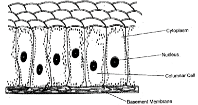
Fig. 8.4.: Columnar epithelium
Function: Being a surface layer columnar epithelium provides protection to underlying tissues. Columnar epithelium of intestine is specialized for the absorption of water and digested food. It is also a component of most glandular epithelia. Some columnar epithelial cells modify into goblet cells and produce mucus.
Table. 8.5. Differences between Squamous Epithelium and Columnar Epithelium
|
|
Squamous Epithelium |
|
Columnar Epithelium |
|
1. |
Its cells are thin and flat. |
1. |
It consists of pillar like cells. |
|
2. |
The nucleus lies in the center which is generally bulged out in squamous epithelial cells. |
2. |
The nucleus lies near the base in columnar epithelial cells. |
|
3. |
The tissue is found in lung alveoli, blood capillaries, Bowman's capsule, skin, ducal cavity etc. |
3. |
The tissue is found in the lining of stomach and intestine, their glands and the covering of epiglottis. |
|
4. |
It functions as selectively permeable barrier, in ultrafiltration and wear and tear. |
4. |
It takes part in absorption, secretion and as protective covering. |
Characteristics: Ciliated epithelium is formed of cuboidal or columnar epithelial cells that bear cilia on their free surfaces. A cilium is very fine, vibratile cytoplasmic projection that arises from a minute basal granule.
Location: Cuboidal ciliated epithelium lines certain parts of urinary tubules of the kidney and sperm ducts. The columnar ciliated epithelium lines the trachea or wind-pipe, bronchi (lungs), kidney tubules and oviducts (fallopian tubes).
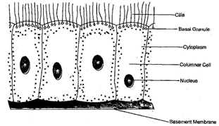
Fig. 8.6: Ciliated columnar epithelium
Function: Ciliated cuboidal and ciliated columnar epithelium both help in the movement of mucus, urine, egg, sperms and cerebrospinal fluid in a particular direction due to rhythmic, beating of the cilia.
Characteristics: These are specialised cuboidal or columnar epithelial cells that forms the glands. Glands are the structure which secrete useful substances, so glands are always epithelial in origin. The glands may be unicellular or multicellular. On the basis of shape of their secretory part multicellular glands may be tubulor or secular.
Location: Glandular epithelium are found in skin, intestine, pancreases, and other glands.
Function: These secrete various type of substances such as sweat, oil, enzymes, hormones etc..

Fig. 8.7. Goblet cell in Columnar Epithelium
9.1. Characteristics: Muscular, tissues are mesodermal in origin. Muscle forming cells are called myoblasts, which form all the muscle of an animal body. Muscle cells are highly elongated and contractile and are called muscle fibres. These can contracts along their longitudinal axis up to one-third or one-half of their length. The cytoplasm of the muscle fibres, called sarcoplasm? is highly contractile. It has numerous mitochondria collectively called sarcosomes, a network of modified SER called sarcoplasmic reticulum numerous glycogen granules to provide energy for its contraction. Sarcoplasm also contain large numbers of fine longitudinal and contractile protein aceous fibrils called myofibrils Muscles are highly specialised for movements and locomotion so these have no power of division and regeneration.
Functions
Types if muscular tissue
There are three major classes of muscular tissues:
Characteristics: Striated muscles are also known as striped or skeletal musclecs which form more than 80% of the mass of soft tissues in a vertebrate body. They are attached to the bones by ten dones and help in the movement of external body parts. Therefore, they are also known as skeletal muscles. They work according to our will, so they are also called voluntary muscles.
The striated muscles are formed of long cylindrical, straight, unbranched fibres or cells which have blunt ends. They are known as muscle fibres. Each fibre is enclosed in a thin but distinct plasma membrane, called sarcolemma. A muscle fibre is about 40 mm in length and 1&-100 pm in diameter. The muscle fibre or cell contains many elongated, flattened nuclei characteristically located towards the periphery near the sarcolemma.
Under light microscope, each muscle fibre appears to be formed of alternate light and dark cross bands. Due to the presence of bands muscles are called striated muscles. The muscle fibres are arranged in bundles and are held together by connective tissue.
Location: striated muscles are found in the muscles of limbs (.e.g., biceps and triceps of arms, face, neck, body wall etc. They are also found in lounge, pharynx, and upper part of oesophagus.
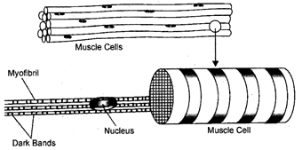
Fig. 9.2: Striated muscles
Functions: Striated muscles help in the movement of body parts and locomotion. They also help in ingestion of food, breathing, blinking of eyes and several voluntary movements of the body.
Characteristics: These are also known as visceral or involuntary muscles. They do not show any stripes across the muscles fibre. Smooth muscles occur as bundles or sheets of elongated fusiform or spindle-shaped fibres or cells which have tapering ends. Each fibre or cell is uninucleate, nucleus is oval and centric. The number and size of mitochondria and glycogen granules are less than those of striated muscle fibres. Sarcoplasmic reticulum is also less developed.
Location: Smooth muscles occur in the walls of visceral organs like alimentary canal, blood vessels, urinary bladder, ureters, genital ducts, so are also called visceral muscles. These are also present in dermis of skin, iris and biliary body of eye.
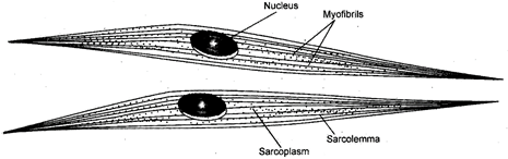
Fig. 9.3. Unstriated (smooth) muscle fibres
Functions
Characteristics: Cardiac muscles resemble both striated and smooth muscle fibres in some characteristics, but also have some peculiar characters of their own. Cardiac muscle fibres resemble striated muscles in being cylindrical, high vascularisation, having more mitochondria and glycogen granules in the sarcoplasm; and having dark and light bands showing similar arrangement of thick myosin and thin actin filaments, covered by sarcolemma. Cardiac muscles resemble smooth muscles in being mostly uninucleate, smaller in size, and in being involuntary.
Cardiac muscles are branched cells and are joined end to end by flat dense, zig-zag junctions called intercalated discs. These allow rapid spread of wave of excitation from one muscle fibre to next throughout the heart. Each fibre or cell contains one or two nuclei, situated in the centre.
The muscle fibres are interconnected by oblique bridges so forming a network.
Location: The cardiac muscle are found exclusively in the wall of heart.
Functions: The cardiac muscles undergo rhythmic contraction and can initiate their own waves of excitation. These contract rapidly like striated muscle fibres but do not get fatigued, so these work with same efficiency throughout life. Contraction and relaxation of cardiac muscle cause pumping of blood out of the heart and into the heart regularly.
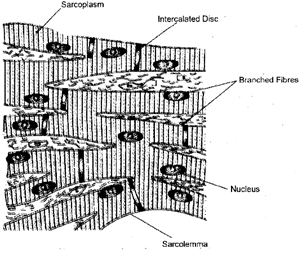
Fig. 9.4.: Cardiac muscles
10.1. Characteristics: Connective tissues provide the structural framework and support to different tissues forming on organ. It is the most abundant and widely distributed tissue. It develops from the embryonic me sperm. It is a fundamental animal tissue having scattered living cells embedded in amorphous, transparent ground substance called matrix that helps in connecting, binding, packing and supporting different structures of the animal body. The matrix and fibres are usually secreted by the cells. It can be jelly-like, fluid or solid. On the basis of nature of matrix, connective tissue is of three types-connective tissue proper (jelly - like matrix), skeletal tissue (solid matrix) and vascular tissue (fluid matrix). The matrix is fibre free in the vascular connective tissue while it is densely mineralised in the bone.
Component of Connective Tissues
Three components are present in the connective tissues:
(i) Intercellular medium (ground substance): If is mainly a mixture of carbohydrates and proteins.
(ii) Connective tissue cells: The cells are of different types:
(a) Fibroblasts: They produce fibres and matrix.
(b) Adipose cells: (Adipocytes or lipocytes): They store fats.
(c) Plasma cells: Synthesize antibodies.
(d) Mast cells: Produce histamine, heparin and serotonin. They are involved in the inflammation of the infected area.
(e) Macrophages: Ingest cell debris and foreign bodies.
(f) Immunocytes: produce antibodies.
(iii) Connective tissues fibres: These are of three types:
Collagen fibres (white fibre)
Elastin fibres (yellow fibres)
Reticular fibres
General Function
Types of Connective Tissues
The connective tissues are of five major types:
Characteristics: if is also known as loose connective tissue. It consists of a transparent, jelly-like (gelatinous) sticky and highly vascular matrix containing numerous fibres and cells and abundant mucin. The fibres are generally two types (a) White collagen fibres, made up of a protein collagen which on boiling with water changes to gelatin and (b) Yellow elastic fibres, made up of (b) protein (a) Collagen fibres provide flexibility and strength whereas elastic fibres provide elasticity.
Location: It most abundant type of connective tissue and is widely distributed in the body. It is found beneath the epithelia of many visceral organs and on the walls of blood vessels.
It joins skin to muscles.
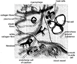
Fig.10.2: Areolar Connective Tissue
Function
Structure: Dense regular connective tissue is a fibrous connective tissue. It is characterized by ordered and densely packed collection of fibres and cells. The fibres are loose and very elastic in nature. Dense regular connective tissue is the principal component of tendons and ligaments.
(i) Tendon: It is a small cord-like dense white fibrous connective tissue which has great strength and limited flexibility. Tendons are formed of parallel bundles of white collagen fibres with rows of flat elongated fibroblasts or tendinocytes in between them. A layer of areolar tissue occurs on the outer surface of tendon. Tendon joins a skeletal muscle to a bone. It helps in moving the bone on contraction and relaxation of the muscle.
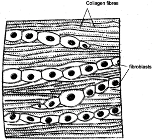
Fig. 10.3.: Dense regular connective tissue
(ii) Ligament: It is also a cord-like dense yellow fibrous connective tissue having considerable strength and high elasticity. A ligament consists of a number of yellow elastin fibres, bundle of white collagen fibres arranged variously and fibroblasts scattered in between the fibres. A ligament joins a bone with another bone. Being elastic, a ligament allows bending and rotation movements over a joint. Sometimes a ligament gets overstretched, producing sprain.
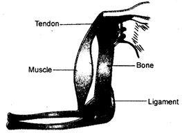
Fig. 10.4.: Attachment of tendons and ligaments
Table 10.5. Differences between Tendon and Ligament
|
|
Tendon |
|
Ligament |
|
1. |
It is tough and less elastic in nature. |
1. |
It is strong but elastic in nature. |
|
2. |
It lacks yellow elastin fibres. |
2. |
It has yellow elastin fibres in good number. |
|
3. |
It contains parallel bundles of white collagen fibres, |
3. |
It contains bundles of while collagen fibres scattered in various directions. |
|
4. |
They occur in rows. |
4. |
They lie scattered. |
|
|
It connects a muscle to a bone. |
|
It joins a bone with another bone. |
Characteristics: It is fat storing tissue and is formed of an aggregation of fat cells or adipocytes. Each adipocyte is rounded or oval in shape and contain a large droplet of fat that almost fills it. The cells have a peripheral cytoplasm with nucleus at one end. Adipose tissue is otherwise similar to areolar tissue in having soft jelly-like matrix. The matrix also contains few fibroblasts, macrophages, collagen fibres and elastic fibres.
Location: The adipose tissue occurs in subcutaneous region, cushion around eyes, kidneys, blood vessels, heart and inside yellow bone marrow.
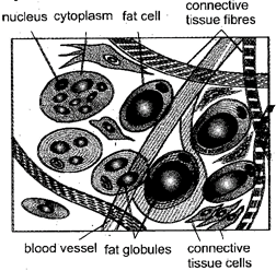
Fig. 10.6.: Adipose tissue
Function
Do You Know
It is also known as supportive connective tissue. It forms the rigid skeleton which supports the vertebrate body, helps in locomotion and provide protection to many vital organs. In skeletal tissue matrix is rigid and the living cells occur in fluid filled space called lacunae. Skeletal tissue is of two types:
(i) Cartilage
(ii) Bone
(i) Cartilage: It is a hard but flexible skeletal tissue consisting of living cells embedded in a matrix rich in protien chondrin. It is a semi solid, flexible, transparent and jelly-like substance which contain 60-80% of water. There are three types of glycos-amino-glycons viz. Hyluronic acid, condroitin sulphate, and keratin sulphate desolved in matrix. Matrix also contains collagen and elastic fibres which provide rigidity to matrix.
The matrix of cartilage is secreted by cells (chondroblasts) that become embeded in the matrix as chondrocytes. These cells are enclosed in lacunae. Usually one or two or four chondrocytes are present in a lacuna. The surface of cartilage is covered by an irregular connective tissue forming the perichondrium except cartilage present on articular surface. Growth of cartilage occurs continuously due to multiplication of chondrocytes by mitosis, deposition of matrix within existing cartilage and from activity of deeper cells of the perichondrium. Blood vessels and nerves are absent in the matrix of cartilage.
Location: Cartilage is found more abundantly in vertebrate embryo because most of the bones forming skeleton of the adult are cartilaginous in the early stage. Otherwise it is found in very few parts of the body. In humans, it occurs at the ends of long bones, the pinnae of ears, the nose, in the trachea as tracheal rings, within intervertebral disc etc. In sharks and ray fishes, the entire skeleton is cartilaginous....
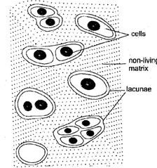
'Fig. 10.7. Cartilage
Functions
(ii) Bone
Characteristics: Bone is a solid, rigid and strong connective tissue in which living cells are embedded in an opaque calcified matrix of ossein. Bone is the hardest tissue of the body which forms the main endoskleton of the vertebrate body.
The Bone consists of a solid matrix with fluid filled lacunae having osteocytes or bone cells.
Unlike cartilage, blood vessels and nerves pass into the interior of bone.
Location: A large number of bones are found in the body which are joined together to form the endoskeleton of animal body.
Function
6. Bones store calcium.
Table 10.8. Difference between Cartilage and Bone
|
|
Cartilage |
|
Bone |
|
1. |
It is tough and elastic |
1. |
It is tough and non-elastic. |
|
2. |
Matrix is formed of only organic matter called chon drin. |
2. |
Matrix is formed of both organic and inorganic matter. Osse in is organic. |
|
3. |
Deposition of mineral is rare. |
3. |
Matrix is vascular. |
|
4. |
Matrix is non vascular i.e. blood supply does not present. |
4.
|
Matrix shows concentric lamellae. |
|
5. |
Matrix is homogenous. |
5. |
Matrix shows concentric lamellae. |
|
6. |
A marrow cavity is a always absent. |
6. |
Marrow cavity is often present. |
|
7. |
Lacunae are without canaliculi |
7. |
Each lacuna gives branched processes, canaliculi. |
|
8. |
Cells occur singly, in two or four. |
8. |
Cells occur singly. |
It is special type of connective tissue which differ from connective tissue in following characters:
(i) The matrix is fluid and fibre free
(ii) The matrix is not secreted by the coils.
(ii) The cells have no power of division.
It circulates in the body and is responsible for the transportation of materials from one place to another within the body. It also plays an important role in defense-mechanism of the body.
Do You Know
Types of Vascular tissue: Fluid connective or vascular tissues are of two types, blood and lymphs Composition of vascular tissues has been shown in following chart.
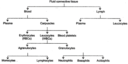
Physical Characters: The blood is red coloured vascular tissue. It is opaque, somewhat sticky and viscous fluid (viscosity = 4.7). Human blood is five times more viscous than distilled water. It is slightly heavier than water (specific gravity = 1.057 in males and 1.053 in females).
It is slightly alkaline in natures (average pH = 7.4). The oxygenated blood is bright red while the deoxygenated blood is purple coloured.
Blood consists of an aqeous mixture of substances in blood plasma in which there are suspended different types of free floating cells (blood corpuscles).
Blood Plasma: It is a pale-straw coloured fluid matrix or medium which constitute about 60% part of the blood. Blood plasma consists of about 90% water and 10% mixture of different types of molecules that enter into the blood at various locations. These substances include- proteins (soluble protein such as albumins, globulins and fibrinogens), glucose, amino acids, lipids, vitamins, enzymes, hormones, urea and uric acid. They belong to the category of nutrients, wastes, hormones and osmoregulators.
Blood Plasma helps in
(i) Transport,
(ii) Retention of fluid in blood
(iii) Maintenance of blood pH,
(iv) body immunity,
(v) Prevention of blood loss,
(vi) Conducting heat to skin for dissipation and
(vii) Uniform distribution of heat all over the body.
Blood Corpuscles
There are three types of blood corpuscles found floating in blood plasma:
3. Blood platelets or thrombocytes
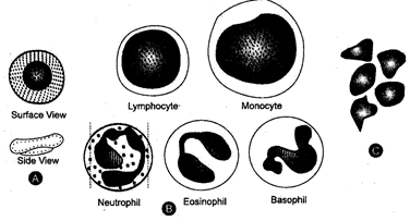
Fig. 10.9: Human blood corpuscles A. Erythrocytes (Red corpuscles); B. Leucocytes
(White blood corpuscles); C. Platelets
Fig. 10.9: Human blood corpuscles A. Erythrocytes (Red corpuscles); B. Leucocytes
(White blood corpuscles); C. Platelets
In all vertebrates except mammals, the erythrocytes are oval, biconvex and nucleated. In mammals, the mature erythrocytes are circular, biconcave and without nucleus A human RBC is about 7.8 im in diameter. Normal RBC count in an adult human male is 5-5.5 million per cubic milimeter of blood. While it is 4.5-5.0 million per cubic milimeter of blood in a normal adult woman. An RBC appears yellow when seen singly but appear red when in bulk due to presence of a colloidal solution of an iron containing pigment haemoglobin in their cytoplasm.
A human RBC is bounded by an elastic and semipenneable plasma-membrane called Donnan's membrane which enables it to squeeze through narrow capillaries. The mature mammalian RBC is also characterised by absence of nucleus, mitochondria and endoplasm reticulum so there is low consumption of oxygen by itself and is primarily concerned with the transport of oxygen from the respiratory organs to the body tissues. RBCs. also contain the enzyme carbonic anhydrase which regulates \[C{{O}_{2}}\]transport. Due to absence of nucleus the RBCs are short lived. Average life span of human RBC is 115-120 days.
These are rounded or amoeboid, nucleated, non-pigmented cells. These are wandering cells and are capable of coming out of blood capillaries by amoeboid movements, called diapedesis, WBCs are larger than RBCs in size. These are much less in number than RBCs. Average WBC count is 7,000 per cubic milimeter of blood. The formation of leucocytes occurs in bone marrow payer's patches, lymph nodes, thymus, spleen etc. WBCs play an important role in the boctyg defence mechanism. On the basis of cytoplasmic granules, the leucocytes are divided into two categories-
Granulocytes which have irregular-shaped nuclei and cytoplasmic granules with specific staining properties. They include neutrophils, basophils and oesinophils. 2. Agranulocytes have no cytoplasmic granules and include monocytes and lymphocytes. Neutrophils engulf and digest disease causing pathogens, eosinophils show allergic response and antihistamine properties and basophils release histamine and heparin. Monocytes engulf bacteria and cellular debris at injured site and lymphocytes secrete antibodies to destroy microbs and also help in healing of injuries.
Table. 10.10. Formed Element of Blood
|
|
Formed Element |
|
Number/Percentage |
|
1. |
Red blood corpuscles (Erythocytes) |
1. |
5-5.5 million/ |
|
2. |
White blood corpuscles (Leucocytes) I. Agranulocytes (i) Lymphocytes (ii) Monocyyes II. Granulocytes (i) Neutrophils (ii) Eosinophils (iii) Basophils |
2. |
6,000-9000/ 20-45% 2-10%
40-75% -1 0-1% |
|
3. |
Platelets |
3. |
200,000-400,000/ |
Table. 10.11. Differences between RBCs and WBCs
|
|
RBCs |
|
WBCs |
|
(i) |
They are red |
(i) |
They are colourless |
|
(ii) |
Size of a RBC is about 7.8 |
(ii) |
Size of WBC varies between 10-20 |
|
(iii) |
They contain haemoglobin |
(iii) |
They do not contain hemoglobin |
|
(iv) |
Nucleus is absent |
(iv) |
Nucleus is present |
|
(v) |
They are biconcave rounded in shape |
(v) |
They are rounded or amoeboid. |
|
(vi) |
Number is more than WBC s. |
(vi) |
Number of less than RBCs |
|
(vii) |
Most cell organelles are absent |
(vii) |
Cell organelles are present |
|
(viii) |
They are only one type |
(viii) |
They are of five type. |
|
(ix) |
They help in transport of oxygen |
(ix) |
They help in defense and immunity |
|
(x) |
Life span is 120 days |
(x) |
Life span is generally short. |
Function of Blood
These also resemble with Leucocytes of blood. These also move from the blood by diapedesis.
Do You Know
Function: The lymph acts as middle man between the blood and the tissue cells as it passes on food and oxygen from blood to tissue cells and handover excretory wastes, hormones, and \[C{{O}_{2}}\]from the body cells to blood. It also keeps the tissues moist. It also transports food fat from the intestine to the venous blood.
Neural or Nervous tissue is ectodermal in origin which receives stimuli and conducts impulses for controlling and coordinating body functions. Brain, spinal cord and nerves are all composed of nervous tissue. Nerve cells or neurons the unit of nervous tissue are highly specialized and excitable cells. Nerve cells or neurons have the ability to receive stimuli from within or outside the body and to conduct impulses to different parts of the body. .The impulses (signals) travels from one neuron to another neuron. Neurons are the longest cells of the body reaching up to a meter in length.
Each neuron is made up of two parts, cell body and neuritis’s Neuritis are of two types, dendrite and axon.
The large dendrites are also called dendrons. Dendrite also possess Nissfs granule and neurofibrils.
Dendrites pick up impulses and transmit the same towards cyton.
3. Axon: It is also known as Nerve fibre. It is long cylindrical process of uniform diameter that arises from the axon hillock of the cyton. It is devoid of nisus granule. It however, contains neurofibrils. It carries impulses away from cyton. Axon is surrounded by a sheath of special connective tissue cells called Schwann cells. The en sheathed axon is called nerve fibre. Axon shows fine branching at its terminal end. Each branch end is a swollen structure called synaptic button. The synaptic buttons of neuron are connected with dendrite branches of an adjacent neuron. Each such junction, in fact has minute gap called synapse. It is ment for the transmission of nerve impulse from one neuron to other.
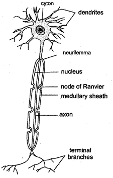
Fig. 11.1: A neuron-a unit of nervous tissue
Function: Nervous tissue pickup sensation such as sight, sound, taste, smell, pain and other stimuli and provides response to all type of stimuli. It exerts control over all body activities.
Nervous tissue or system coordinates the functioning of different body parts.
Table 11.2. Differences between Dendrite and Axon
|
|
Dendrite |
|
Axon |
|
(i) |
It is a short tapering process of neuron. |
(i) |
It is long uniformly thickened fibre-like process. |
|
(ii) |
Dendrite does not have a sheath, |
(ii) |
It may be unsheathed. |
|
(iii) |
Both Nissl?s granule and neurofibrils are present, |
(iii) |
Nissl?s granule absent. Neurofibrils, are however, present. |
|
(iv) |
It carries impulses towards the cell body |
(iv) |
It carries impulses away from the cell body. |


You need to login to perform this action.
You will be redirected in
3 sec