Excretory System Of Man
Category : 11th Class
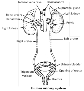
Mammalian (human) urinary system consists of a pair of kidneys, a pair of ureter, a urinary bladder and a urethra.
(1) Kidneys : The kidneys are dark-red, bean-shaped organs about 11 cm long, 5 cm wide and 3 cm thick, each weight about 150 gm in an adult male and about 135 gm in adult female. They are placed against the back wall of the abdominal cavity just below the diaphragm, one on either side opposite the last thoracic and first three lumber vertebrae. The 11th and 12th pairs of ribs protect them.
The kidneys are covered by peritoneum on the front (ventral) side only. Thus, they are retroperitoneal. The right kidney is attached more anterior than the left in rabbit. This asymmetry is just the reverse of that found in man.
In man left kidney occurs at a slightly higher level than the right one, because right side has prominent right liver lobe. In rabbit the condition is little differ due to quadropedilism i.e. left kidney is in normal position while the right kidney shift ahead to provide place for stomach below it.
In mammals, the kidney is concavo convex. The center of concave inner surface is called as hilum or hilus which gives out a ureter. From this hilus surface the renal artery enters into the kidney, the renal vein comes out and the renal nerves enter into the kidney.
(i) Structure of kidney : The kidneys are metanephric in mammals. The kidney is divisible into two parts outer-cortex and inner-medulla. Three layers of tissue surround each kidney.
(a) The innermost, renal capsule made up of fibrous connective tissue.
(b) The intermediate layer, adipose capsule of fatty tissue.
(c) The outermost, renal fascia of dense connective tissue.
Renal pyramids or medullary pyramids : The medulla is subdivided into 8 to 18 conical masses - the renal pyramid, each having broad base towards the cortex and a narrow end called renal papilla towards the pelvis.
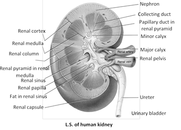
Path of urinary drainage : Collecting duct\[\to \]Papillary duct in renal pyramid\[\to \]Minor calyx\[\to \]Major calyx\[\to \]Renal pelvis\[\to \]Ureter\[\to \]Urinary bladder
Renal columns of bertini : Between the pyramids, the cortex extends into the medulla or renal columns of bertini.
Calyx : Each renal papilla projects into the cavity of a minor calyx, minor calyx join to form major calyx. The major calyx open into a wide funnel like structure, the pelvis. The latter leads into the ureter. In rabbit, the pelvis is unbranched hence, it is without calyx.
In frog ventral surface of each kidney has many ciliated funnels called nephrostomes. They drain wastes from body cavity (coelom) and connect to renal veins in frog or to uriniferous tubules in tadpoles.
(ii) Histology of kidney : Histologically a kidney is made of innumerable thin, long, much convoluted tubular units called uriniferous tubule or nephron.
Nephron is the structural and functional unit of kidney. One human kidney may contain about one million (10 lac nephron) nephron (In rabbit each kidney bear about 2 lac nephron). In frog each kidney bears about 2 thousand nephron.
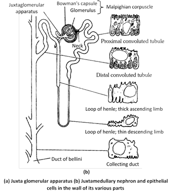
(a) Structure of nephron : A nephron or uriniferous tubules consist of two parts :
Malpighian body / Renal Corpuscles : The proximal end of each nephron forms a blind or closed, enlarged and double walled cup, the Bowman's capsules in the cortex. (name Bowman's capsule is based on english physiologist and histologist William Bowman).
Each capsule contains a network of blood capillaries the glomerulus which receives blood through afferent arteriole and the blood comes out through the efferent arteriole .The diameter of the efferent arteriole is comparatively lesser. (Bowman's capsule and glomerulus receives about 20 – 25% of the cardiac out put (blood) at rest.
The composite structure of Bowman's capsule and glomerulus is known as Malpighian body or Malpighian corpuscles after the Italian microscopist Marcello Malpighi.
Tubule : The tubule is differentiated in to 3 parts P.C.T., Henle's loop and D.C.T.
The Bowman's capsule opens into a proximal convoluted tubule (P.C.T.) the anterior part of the P.C.T. is more coiled where as its posterior part is almost straight. The P.C.T. opens into a Henle's loop. The Henle's loop is a U- shaped structure or makes hair pin turn, which has a distinct descending limb and an ascending limb. The ascending limb opens in to the distal convoluted tubule. The D.C.T. is a coiled structure. Many D.C.T. unit to form a collecting duct. The collecting ducts of one pyramid unit to form a duct of Bellini. The duct of Bellini lead into the pelvis part.
(b) Arrangement of nephron : The malpighian body and most of the P.C.T. and D.C.T. are situated in the cortex. Henle's loop and collecting ducts are found in the medulla.
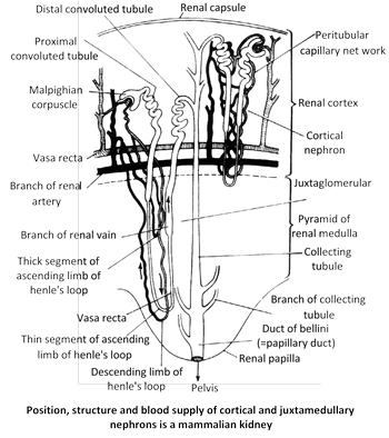
Vasa recta : The efferent arteriole of juxta-glomerular nephron forms a peritubular capillary system around the Henle's loop which is called vasa recta. Each of the vasa recta makes U turn at the inner most part of the medulla and return to the venous circulation near the junction of medulla and cortex. The efferent arteriole and peritubular capillaries technically constitute a renal portal system. In all amniotes as reptiles, birds and mammals have a this renal portal system of efferent arteriole and peritubular capillaries.
(c) Types of nephron : Nephrons are of two types cortical and juxtamedullary, with regard to their location in the kidney. The cortical nephrons form about 80% to 85% of total nephron. They lie in the renal cortex and have very short loops of Henle that extend only little into the medulla.
Remaining 15 - 20% are juxta medullary nephron have their Bowman's capsule close to (Juxta) the junction of the cortex and the medulla and have very long loops of Henle, extending deep into the medulla. This type of nephron is present in only birds and mammals. The cortical nephrons control the plasma volume when water supply is normal. The juxtamedullary nephrons regulate the plasma volume when water is in short supply (In advarse condition).
Differences between cortical and Juxtamedullary nephrons
|
S.No. |
Cortical Nephrons |
Juxtamedullary Nephron |
|
1. |
Form 80 - 85% of total nephrons. |
Form only 15 - 20% of total nephrons. |
|
2 |
Are small in size. |
Are large in size. |
|
3. |
Lie mainly in the renal cortex. |
Have Bowman's capsules in the cortex near its junction with the medulla. |
|
4 |
Henle's loops are very short and extend only a little into the medulla |
Henle's loop are very long and extend deep into the medulla. |
|
5. |
Control plasma volume when water supply is normal. |
Control plasma volume when water supply is short. |
|
6. |
Glomeruli in superficial region of cortex |
Glomeruli deep in cortex |
|
7. |
Blood supply from peritubular capillaries only. |
Blood supply from both peritubular capillaries, and vasa recta. |
(d) Histology of nephron
Glomerulus : Glomerulus is a network of up to 50 parallel branching and anastomosing capillaries covered by endothelium, basement membrane and epithelium made of podocytes which has slit pores that restrict passage of colloids. However, small molecules and water can easily pass through them in to the P.C.T.
Bowman's capsule : The podocytes forming the inner wall of the Bowman's capsule have gaps (about 25 nm wide) the slit pores.
The outer wall of the Bowman's capsule consists of unspecialized squamous epithelium (flattened).
Proximal convoluted tube : P.C.T. is made up of simple cuboidal epithelium . It has microvilli so it is also known as brush border epithelium. P.C.T. is most important site for selective reabsorption.
Loop of Henle : The epithelium of descending limb of loop of Henle is very thin and composed of squamous epithelium and ascending limb is made up of two parts. First is thin ascending limb lined by squamous epithelium and second thick ascending limb lined by cuboidal epithelium. The ascending limb is impermeable to water and permeable to \[NaCl.\]
Distal convoluted tube : D.C.T. is made up of cuboidal epithelium which is glandular in nature.
Collecting ducts : The collecting ducts are lined by cuboidal epithelium in different regions. At intervals, the cuboidal cells are ciliated.
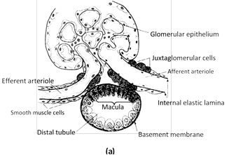
Juxta-glomerular apparatus : This specialized cellular apparatus is located where the distal convoluted tubule passes close to the Bowman's capsule and afferent arteriole. Cells of the D.C.T. epithelium in contact with afferent arteriole are denser than other epithelial cells known as maculla densa. Maculla densa has special Lacis cell or Polkisson's cell. These cells secrete renin hormone that modulate blood pressure and thus renal blood flow and G.F.R. are regulated.
Origin and types of kidneys in different vertebrate
Kidney tubules (nephrons) arise in the embryo in a linear series from a special part of mesoderm called mesomeare or nephrotome.
Number, complexity and arrangement of Nephrons are differ in different groups of vertebrates. A nephron is differentiated into three parts – peritoneal funnel, tubule and malpighian body. Peritonial funnel (nephrostome) are normally present in embryos and larvae and considered as vestigeal organ of hypothetical primitive kidneys.
(1) Archeonephros kidney : Archeonephros is the name given to the hypothetical primitive kidney of ancestral vertebrate. It is also called as holonephros or complete kidney. (It extended entire length of coelom) It tubules are segmentally arragned and nephrostome is present. Glomerulus is external (without capsule). It duct is called as archeonephric duct. Ex. Larva of myxine and some apodan amphibians.
Modern vertebrates exhibits three different kinds of adult kidney Pronephros, Mesonephros and Metanephros.
(i) Pronephros : It originates from the anterior part of the nephrotome. It is also termed head kidney due to its anterior position. There are only 3 pronephrine tubule (nephron) in frog embryo, 7 in human embryo, and about 12 in chick embryo which are segmentary arranged. Nephrostome present, glomerulus is external and unite to form glomus in some cases. Duct is pronephric duct or mullerian duct. A pair of pronephros appear in all vertebrate embryos but they becomes functions kidneys in adult myxine and embryos of all anamniotes (fish, amphibian). This kidney found as transitory kidney in all vertebrates embryos.
(ii) Mesonephros : It originates from the middle part of the nephrotome. Duct is mesonephric or Wolffian duct. Nephrostome is absent except some embryos of anamniotes. Example – In amniotes (reptiles, birds and mammals) mesonephros is functional only in the embryos, replaced by metanephros in the adult. In anamniotes (fishes and amphibian) mesonephros is functional in both embryo as well as adults. Also found in adult petromyzon.
(iii) Metanephros : It originates from the posterior part of the nephrotome. When metanephric tubules develop, all the mesonephric tubules disappear except those associated with the testes in male and forming vasa efferentia. Nephrostome absent. A thin, U-shaped loop of Henle forms between P.C.T. and D.C.T. which is incomplete in Reptiles and Birds and well developed in mammals. Duct is metanephric or ureter. Reproductive duct is separate. The kidney is highly compact which possesses innumerable nephrons. Example – All amniotes – Reptile, Birds and mammal.
(2) Ureters : From the hilum of each kidney emerges a whitish tube the ureter. The ureters are about 28 cm long. Their wall consists of transitional epithelium surrounded by a layer of muscle fibres. Openings of the two ureters in the bladder are separate, but closely placed. These are oblique, so that the urine cannot regurgitate into the ureters when the bladder contracts. Peristalsis of ureters also cheeks regurgitation of urine. Like kidneys, the ureters are retroperitoneal.
(3) Urinary bladder and Urethra : The urinary bladder is pear-shaped hollow muscular organ situated in pelvic cavity, which is made up of smooth and involuntary muscles. The muscles is also known as detrusor muscles (muscles that has the action of expelling a substance). The lower part or neck of the bladder leads into the urethra. There is a smooth triangular area, called trigonium vesicae. The lumen of the urinary bladder is lined by transition epithelium which has great power of streaching. The neck of bladder is guarded by two sphincters, inner is involuntary controlled by spinal reflex and outer is voluntary controlled by cerebral cortex. A person feels the sensation of micturation when the quantity of urine in the bladder is about 300 c.c. The average capacity of urinary bladder is 700 - 800 ml. In general, urinary bladder capacity is smaller in females because the uterus occupies the space just superior to bladder. Mucosa of bladder with folds called rugae. (rugae also present is stomach and vagina).
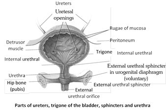
(4) Urethra : The urinary bladder leads into the urethra. In a female, it is quite short, only about 3 to 5 cm long, and carries only urine. It opens by urethral orifice, or urinary aperture in the vulva infront of the veginal or genital aperture. In a male urethra is much longer, about 20 cm and carries urine as well as spermatic fluid. It passes through the prostate gland and the penis. It opens out at the tip of the penis by urinogenital aperture. In males the epithelium of spongy urethra is stratified or pseudostratified columnar epithelia, except near external urethral orifice, which is non keratinized stratified squamous epithelia. The prostatic urethra lined by transitional epithelia, while membranous urethra lined by pseudostratified columnar.
Differences between male and female urethra
|
S. No. |
Male urethra |
Female urethra |
|
1. |
It is about 20 cm long. |
It is just \[35\text{ }cm\]long. |
|
2. |
It has 3 regions : prostatic urethra \[(34\text{ }cm),\] membranous (1 cm) and penile (15 cm) |
It is not differentiated into regions. |
|
3. |
It opens out at the tip of the penis by urinogenital aperture. |
It opens into the vulva by urinary aperture. |
|
4. |
It carries urine as well as semen to the exterior. |
It carries only urine to the exterior. |
|
5. |
It has 2 sphincters. |
It has a single sphincter. |


You need to login to perform this action.
You will be redirected in
3 sec

