Some Representative Protozoan Protists
Category : 11th Class
![]()
General characters
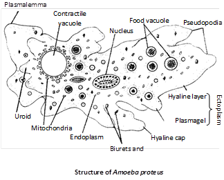
(1) Amoeba belongs to the class Sarcodina or Rhizopoda of the phylum protozoa. It is discovered by Russel Von Rosenhoff in 1755.
(2) The most common species of Amoeba proteus. Proteus is the name of the mythical sea god who could change shape.
(3) Amoeba is cultured in laboratory by Hay infusion method.
(4) Body is covered by plasmalemma. It is a trilaminar and selectively permeable membrane. Plasmalemma is excretory, ammonia diffuses out through it. It is also respiratory diffusion of oxygen and carbon dioxide takes place through it.
(5) The body bears a member of temporary and blunt pseudopodia. The type of pseudopoium found in Amoeba proteus is lobopodium. Pseudopodia are composed of both ectoplasm and endoplasm.
(6) Pseudopodium at its forward end gets its from consistency by hyaline cap which is made of ectoplasm.
(7) Pseudopodia in Amoeba are meant for feeding and locomotion.
(8) Pseudopodia are found in Amoeba and leucocyte of higher animals.
(9) Cytoplasm is differentiated into outer ectoplasm and inner endoplasm. Endoplasm is divided into outer plasma gel and inner plasma sol.
(10) Locomotion of Amoeba is known as amoeboid movement. Sol gel theory of amoeboid movement was first given by Hyman supported by Pantin and Mast. According to this theory amoeboid locomotion is due to change in the velocity of cytoplasm.
Theories of Amoeboid Movement
|
Surface tension theory |
Berthold |
(1886) |
|
Rolling movement theory |
Jennings |
(1904) |
|
Walking movement theory |
Dellinger |
(1906) |
|
Sol-gel theory |
Hyman |
(1917) |
|
Folding and unfolding theory |
Goldacre and Lorch |
(1950) |
|
Contraction-hydraulic theory |
Rinaldi and Jahn |
(1963) |
(11) Sol gel conditions are due to contraction and relaxation of long chains of proteins.
(12) Amoeba contains a nucleus, a contractile vacuole, a number of food vacuoles and other cell organelles.
(13) Endoplasm of Amoeba in the posterior part contains a single clear rounded and pulsating contractile vacuole. Contractile vacuole is concerned with osmoregulation, i.e., removal of excess of water.
(14) Vacuole is found only in fresh water forms. It is absent in marine and parasitic forms.
(15) If an Amoeba is placed in distilled water its contractile vacuole works faster while it is placed in salt water, its contractile vacuole will disappear.
(16) Contractile vacuole of Amoeba is analogous (similar in function) to uriniferous tubules of frog.
(17) Food of Amoeba consists of bacteria, diatoms, small protozoa and algae.
(18) The process of ingesting solid food is phagocytosis, Amoeba ingest food by import, circumfluence, circumvallation or invagination.
(19) Import involves passive sinking of food into body by rupture of plasmalemma, e.g., Ingestion of algae.
(20) Circumfluence is the ingestion of less active or motionless organisms like bacteria.
(21) Circumvallation is the engulfment of active prey like ciliate or flagellate.
(22) Digestion in Amoeba is intracellular. Amoeba secretes digestive enzymes for hydrolysing starch, protein, fat etc.
(23) Food vacuole of Amoeba is analogous to the alimentary canal of an animal or gastro vascular cavity of Hydra. The contents of food vacuole in Amoeba first becomes acidic then alkaline.
(24) Egestion of undigested food in Amoeba takes place through a temporary rupture of the surface membrane.
(25) Amoeba responds to environmental conditions. Response to the stimuli is called taxis. Different taxis are thermotaxis (temperature) phototaxis (light), thigmotaxis (touch), chemotaxis (chemicals), galvanotaxis (electric current), geotaxis (gravity) and rheotaxis (water current).
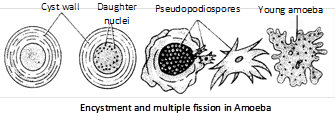
(26) Normal method of asexual reproduction is binary fission. Binary fission is a process of mitosis. It takes place when food is abundant and temperature is suitable.
(27) Multiple fission or sporulation takes place during unfavorable conditions after encystment. There are three layers of cysts.
(28) Lack of oxygen and food induces encystment product of multiple fission are called ?Amoebulae?.
(29) Amoeba regenerates from nucleated bits.
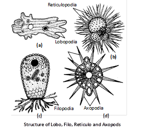
Pseudopodia : These are found in those forms which do not possess a definite pellicle. According to size, structure, and shape pseudopodia may be of different types as :
Lobopods : These are short, blunt and thick finger like out growth of ectoplasm with an axial core of endoplasm e.g., Amoeba, Arcella etc.
Filopods : They are cylindrical thread like, formed entirely of ectoplasm and radiate from the body in all directions e.g., Euglypha, Radiolaria.
Reticulopods : They are filamentous which form a network of pods e.g., Polystomella, Chlamydophrys.
Axopods : These are long stiff semi-transparent extensions of cytoplasm with pointed distal ends e.g., Actinophrys.
![]()
General characters
(1) Entamoeba histolytica is a parasitic and pathogenic protozoan protists which resides in the upper part of large intestine in human beings. It causes amoebic dysentery or amoebiasis.
(2) Lamble (1859) discovered E. histolytica. Friedrick Losch, a Russian Zoologist, discovered its pathogenic nature in 1875.
(3) It has two forms, adult trophozoite or magna, pathogenic form found in the mucosa and sub-mucosa of intestine forming ulcers and minuta, nonpathogenic form found in the lumen of intestine.
(4) Entamoeba has no contractile vacuole.
(5) Trophozoite of Entamoeba reproduces by binary fission. Minuta form encysts. A mature cyst is called quadrinucleate cyst. It has four nuclei and two chromatoid bodies.
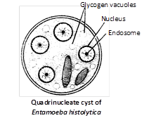
(6) Quadrinucleate cyst is the infective stage. Infection is oral through contaminated food and water.
(7) The reserve food material in cyst of E. histolytica is glycogen. A single cyst of E. histolytica produces eight amoebulae.
(8) It damages instestinal mucosa by secreting an enzyme-histolysin.
![]()
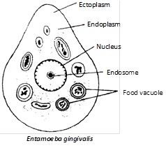
Entamoeba gingivalis is a parasite of human teeth, found in the abscesses of gum and in pus pockets of pyorrhoea, bleeding gums. It increase pyorrhoea disease but does not cause it. Pyorrhoea is caused by Trichomonastinax. Adult is called trophozoite and has 2-3 pseudopodia. It feeds on WBCs, bacteria and pus cells. Cyst is not formed in E. histolytica and infection occurs by direct contract like kissing.
![]()
General characters
(1) Trypanosoma gambiense is the parasite zooflagellate which causes one of the deadliest ailments in human beings called African sleeping sickness or Trypanosomiasis. It was discovered by Frode in 1901.
(2) Trypanosoma is usually found in the blood of vertebrates, finally invading cerebrospinal fluid.
(3) Trypanosoma is an endoparasite, blood parasite, extra cellular parasite.
(4) Trypanosoma has a nucleus, a flagellum, undulating membrane, blepharoplast (basal granule) and kinetoplast. The flagellum arises from the posterior end and runs anteriorly with undulating membrane.
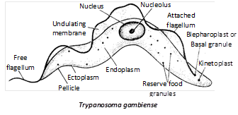
(5) Trypanosoma reproduces asexually by longitudinal binary fission. It does not form cysts.
(6) Trypanosoma is polymorphic and has four forms: Leishmania, Leptomonad, Crithidial and Trypanosomal (= Metacyclic) stages.
(7) Trypanosoma is digenetic, it completes its life cycle in two hosts. The primary or principal or definite host is man and the intermediate or secondary host or vector is the insect, tse-tse fly or bug.
(8) Three important species of Trypanosoma for which man is host are : Trypanosoma gambiense, T. rhodesiensi and T. cruzi.
(9) The chief vector host of T. gambiense transmitting the disease from one man to another is the tse-tse fly, Glossina palpalis. Occasionally, Glossina tachinoides also act as a vector.
(10) T. rhodesiensi causes Rhodesian trypanosomiasis, it is confined to east central parts of Africa, particularly Rhodesia. The insect vectors for T. rhodesiense are tse-tse flies mainly Glossina morsitans and G. pallidipes.
(11) T. cruzi is the causative agent of South American trypanosomiasis or Chaga?s disease T. cruzi is transmitted by bugs like Triatoma and Panstrongylus. Symptoms of Chaga?s disease are fever, diarrhoea, anaemia and enlargement of lymphoid glands etc.
![]()
They are spore forming parasitic protists which lack locomotory structure and contractile vacuoles. The body is covered by pellicle or cuticle.
ystematic position
|
Phylum |
Protozoa |
|
Sub-phylum |
Plasmodroma |
|
Class |
Sporozoa |
|
Sub-class |
Telosporidia |
|
Order |
Haemosporidia |
|
Genus |
Plasmodium |
|
species |
vivax |
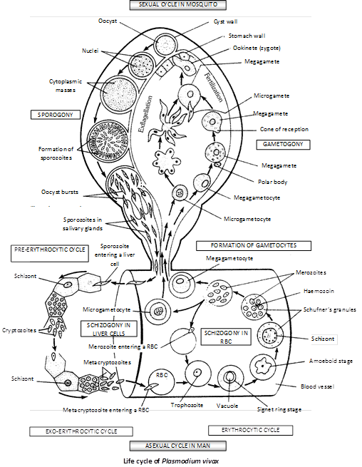
History : The term malaria was coined by Mucculoch in 1827.
Lancisi first suspected a relationship between malaria and mosquito.
Laveran (1880) discovered that malaria is caused by a protozoan parasite, Plasmodium vivax.
Sir Ronald Ross was (1896) the first to observe oocytes of Plasmodium in female Anopheles.
Gassi (1896) was the first to describe the life cycle of Plasmodium in Anopheles.
Host : It is digenetic i.e., life cycle completed on two hosts -
(1) Man (medically primary but biologically secondary host)
(2) Female Anopheles (medically secondary but biologically primary host).
Life cycle : During life cycle two important phases are present.
(1) Endogenous or Asexual phase : passes in man.
(2) Exogenous or Sexual phase : passes in female Anopheles mosquito.
In Man (Schizogony)
Infective stage : It is spindle shaped sporozoite introduced by female Anopheles along with saliva (for anticoagulant anophilin) during blood sucking. Sporozoite enters liver cells. In liver they produce several stages.
Pre-erythrocytic stage : Each sporozoite enters inside the liver cell and becomes spherical and termed as cryptozoite. It undergoes a pre-erythrocytic cycle completed in 10 days and multiplies asexually by schizogony. After rupturing schizont numerous cryptomerozoites are liberated.
Exo-erythrocytic stage : Cryptomerozoites enter in new liver cells and reproduce asexually to give rise to a large number of meta-cryptomerozoities, few are smaller in size and called micro-metacryptomerozoites.
Erythrocytic stage : Micro metacryptomerozoites enter into the blood stream and each enters the RBCs and assumes rounded disc like shape with single nucleus. It develops a vacuole which gives a ring like appearance. This stage is known as signet ring stage. During further development the vacuole is lost and parasite feeds on the cytoplasm of R.B.C. This stage is known as amoeboid stage or trophozoite stage. The matured trophozoite develops to become a schizont. It multiplies asexually by erythrocytic schizogony.
The schizont withdraws its pseudopodia and changes into rounded form. The nucleus divides into large number of nuclei which are arranged on the periphery and collect a little amount of cytoplasm around each. In this way merozoites are produced. This stage is known as Rosette stage. Merozoites are liberated into the plasma. New merozoites again enter fresh RBCs and repeat the erythrocytic cycle to produce large number of merozoites. Haemozoin liberated in blood plasma.
• Paroxysm : Actual attack of malaria occurs after a few initial erythrocytic cycles due to accumulation of haemozoin and toxins in blood. It involves rigour, febrile and defervescent stages.
Formation of gametes : Some merozoites do not enter schizogony but instead form gametocytes in RBCs. Gametocytes are of two types :
(1) Macro-gametocytes : These are female cells which are large in size, laden with food material and having nucleus at one end.
(2) Micro-gametocytes : These are male cells, small in structure and nucleus placed centrally.
In Mosquito (Sporogony)
When female Anopheles bites a patient of malaria, the parasites enter the alimentary canal of the insect. In the stomach of mosquito the asexual forms are digested and only gametocytes survive. Gametocytes are released in blood plasma in large numbers during mid night. They die by morning. Megagametocyte produces only one megagamete. Microgametocyte produces 4-8 motile microgametes by exflagelation.
Fertilization : Both mega and microgamete fuse, form zygote which narrows to becomes ookinete. Ookinete pierces the stomach wall and forms a cyst on its outer surface.
Sporogony : Oocyst undergoes a process of sporogony. The irregular cells thus formed are known as sporoblasts. The nucleus of sporoblast divides several times to give rise to daughter nuclei which migrate in the projections of sporoblast.
These minute projections change into the form of sporozoites. After maturity oocyst burst out and numerous sporozoites are liberated into the body cavity of mosquito. The sporozoites move towards salivary glands and bore into them. In this way they are ready for transmission.
Control of malaria
(1) Prevention from infection is called prophylaxis.
(2) Quinine is obtained from bark of Cinchona (discovered in Peru) which is most commonly used against malaria.
(3) Peludrine, atabrine, camoquinine, Chloroquinine are few other drugs effective against malaria.
(4) M. E. P. is Malaria Eradication Programme.
(5) G.B.P. is General Blood Picutre i.e., blood film made for the test of malaria.
|
Name |
P. vivax |
P. malariae |
P.ovale |
P. falciparum |
|
1. Site |
Liver and RBC of man |
Liver and RBC of man |
Liver and RBC of man |
Liver and RBC of man |
|
2. Distribution |
Tropical countries subtropical |
Tropical countries subtropical |
West Africa and South America |
Tropical countries |
|
3. Duration of exoerythrocytic cycle or prepatent period |
8 days |
14-15 days |
9 days |
5 days |
|
4. Duration of erythrocytic cycle |
48 hours |
72 hours |
48 hours |
48 hours |
|
5. Incubation period |
12-14 days |
20-24 days |
14 days |
12 days |
|
6. Duration of sexual cycle |
10 days |
26-28 days |
16 days |
10-12 days |
|
7. Disease (Type of malaria fever) |
Benign and tertian malaria / relapse malaria |
Quartan malaria clinical malaria |
Ovale and tertian recuris every third day |
Malignant tertian malaria Pernicious malaria Estivoautmnal malaria |
|
8. Pigment and color |
Schuffner?s granules Yellow / brown |
Zeiman?s dot Dark brown |
Schuffner?s dots Dark brown |
Maurer?s dots Dark green |
![]()
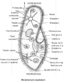
(1) Paramecium is a holotrichous ciliate protozoan. It is discovered by Hill in 1752.
(2) Paramecium is free-living and aquatic form. In laboratory, Paramecium is cultured by ?Hay-infusion method?.
(3) Paramecium is commonly called as ?Slipper animalcule?. Body is distinguished into an oral or ventral surface and an aboral or dorsal surface.
(4) Body is covered with a thin, firm, flexible membrane called pellicle. Entire body surface is covered by numerous cilia, the locomotory organelles. Cilia in the posterior end are longer called caudal tuft. Each cilium arises from a basal granule or kinetosome. Paramecium has infraciliary and neuromotor system to co-ordinate ciliary beat.
(5) Trichocysts are peculiar bottle-shaped organelles present in the ectoplasm of Paramecium. Trichocysts are the organelles of offence and defence.
(6) Paramecium is heterokaryotic (dimorphic nuclei) i.e., macronucleus and micronucleus. Macronucleus is one, large, kidney shaped, controls vegetative functions (metabolism). Micronuclei, one (P. caudatum), two (P aurelia) and several (P. multimicronucleatum) are only concerned with reproduction.
(7) Oral apparatus consists of cytopharynx and cytostome (mouth), cytopyge or cytoproct (anus). Nutrition or food intake in paramecium is holozoic. Paramecium is a filter feeder and feeds on small protozoa, unicellular plants (algae), diatoms, yeast etc. and small bits of animals and vegetables. Most favourite food is Tetrahymena, another ciliate protozoa.
(8) Digestion in paramecium is intracellular. Food vacuole constantly moves along a definite courses (cyclosis) within streaming endoplasm. Food vacuole is digested in the cell body in acidic to alkaline media. Egestion of undigested food takes place through cytopyge or cytoproct, a temporary formed anus.
(9) Paramecium reproduces by transverse binary fission and nuclear reorganisation. Binary fission occurs during favourable condition. In this process, macronucleus divides amitotically and micronucleus mitotically.
(10) Paramecium undergoes several kinds of nuclear reorganisation such as conjugation, autogamy, cytogamy, endomixis and hemimixis. Nuclear reoganisation takes place for rejuvenation.
(11) Conjugation occurs between two mating types of same species of Paramecium. It is a modified form of cross fertilization. Each conjugant produces a female stationary and a male migratory nucleus by three successive divisions of micronucleus. They are called pronuclei.
(12) Synkaryon is the diploid nucleus formed by the fusion of stationary and migratory nuclei in conjugant. Synkaryon divides thrice to form eight nuclei. At the end of the conjugation each Paramecium (exconjugant) produces four daughter Paramecia.
(13) Autogamy is a process of self fertilization. It occurs in a single animal of P. aurelia. Autogamy results in the production of two daughter paramecia from each.
(14) Cytogamy occurs in P.caudatum. The two cytogamonts do not exchange their male pronuclei. Endomixis occurs in P.aurelia. It is an asexual reproduction. Hemimixis is the process of purification act on the part of meganucleus.
(15) Paramecium has kappa, Lambda, Mu and Pi particles in cytoplasm. They differentiate paramecia between sensitive and killer forms.


You need to login to perform this action.
You will be redirected in
3 sec

