Vascular Tissues
Category : 11th Class
It is a mobile connective tissue derived from mesoderm which consists of fibre-free fluid matrix and specialised living cells that are not formed in situ, can neither divide nor secrete matrix. Vascular tissue regularly circulates in the body, takes part in transport of material and performs such activities as scavenging healing of wounds and defence against pathogens. Vascular tissue is of two types, blood and lymph,
Blood
In chordates, and in annelids amongst the non chordates, the blood is a red and opaque fluid of salty taste and peculiar smell. It is a little heavier than water. The study of blood is called haematology. It is red coloured liquid connective tissue which originates from the mesoderm. It reaches into the various organs through the blood vessels and transports various chemical substances between different tissues. During embryonic state, the blood is mainly formed in the liver but little blood is also formed in the spleen and ribs. In adults, the blood is formed in the red bone marrow. The blood formation is called as haemopoiesis.
Viscosity \[\,4.7,\text{ }{{p}^{H}}7.4\]
Specific gravity \[\text{ }10.4\text{ }1.07\]
Volume \[\text{ }5-6\text{ }litre/70\text{ }Kg~\,or\text{ }1/{{13}^{th}}\] part of total body weight
Plasma
It constitutes about 5% of body weight. It represents matrix of blood. Plasma is slightly alkaline and transparent. It forms 55-60% by volume of blood. Plasma contains : Water \[(91-92%),\] Solid \[(8-9%).\]Plasma solid part consists of organic (7%) and inorganic (1%) substances which are as follows :
Organic constituents of plasma : Some are its own constituents, while others are those which are transported by it. All these are divisible into following categories :
(1) Plasma proteins : Protein constitute about 7% part of plasma and remain in it as colloid particles. These mainly include albumins, globulins, prothrombin and fibrinogen.
Globulins are mainly formed by plasma cells in lymphoid organs. Other plasma proteins are mainly formed in liver. These render the plasma viscous, and maintain its osmotic pressure (7.5 atmospheric) and pH. Prothrombin and Fibrinogen are essential for blood clotting. Albumins are mainly responsible for maintaining osmotic pressure in plasma and for osmoregulation in cells and tissue fluids. Globulins help in osmoregulation and transport of proteins and other substances, but most globulins are immunoglobulins, which act as antibodies, destroying harmful bacteria, virus and toxins in blood and tissue fluids. Some proteins, acting as enzymes, also occur in the plasma.
(2) Digested nutrients : These include glucose, fats, fatty acids, phospholipids, cholesterol, nucleosides, amino acids, vitamins etc. These are the supplied by the blood to all cells of body.
(3) Excretory substances : These chiefly include ammonia collected by blood from body cells and urea, uric acid, creatine, creatinine etc., collected mainly from the liver and transported to kidneys for excretion.
(4) Hormones : These are secreted and released in blood by endocrine glands.
(5) Dissolved gases : Each 100 ml. of water of blood plasma contains about \[0.29\text{ }ml\]of \[{{O}_{2}},\text{ }5\text{ }ml.\] of \[C{{O}_{2}}\] and 0.5 ml of nitrogen dissolved in it.
(6) Defence compounds : Certain immunoglobulins or antibodies and some other substances, such as lysozyme (a polysaccharide) and properdin (a large protein) always occur in the plasma. These serve to destroy bacteria, viruses and toxic substances that may enter into the blood from outside, or from body tissues.
(7) Anticoagulant : Mast cells of connective tissues continuously release, in blood plasma, a conjugated polysaccharide, named heparin. The latter serves to prevent coagulation of blood while it is flowing in intact blood vessels.
Inorganic constituents of plasma : Chloride and bicarbonate salts of sodium are the main inorganic constituents. Traces of other salts, like phosphates, bicarbonates, sulphates and iodides of calcium, magnesium and potassium are also found. All salts constitute about 1% of plasma. These remain as ions (electrolytes) and maintain the alkalinity of plasma. A balanced quantity of salt ions in the plasma is essential for proper functioning of nervous system, muscles and other tissues.
Blood corpuscles
Blood corpuscles form 40-50% of the blood and are of three types viz. Red blood corpuscles, white blood corpuscles and platelets.
(1) Red blood corpuscles (RBCs or Erythrocytes) : These occur only in vertebrates and are the most abundant (99%) of blood corpuscles, imparting the characteristic red colour to the blood. The shape, size and structure of RBCs vary in different types of vertebrates, but their function is the same in all, namely to transport respiratory gases, especially the oxygen \[({{O}_{2}}).\]
RBCs of frog : Amphibian RBCs are largest amongst the vertebrates. Those of Amphiuma and Proteus are largest amongst amphibians about \[82\,\,\mu m.\] These are flattened and oval, disclike, but slightly biconvex due to a large oval and centrally-placed nucleus.
RBCs of mammals : Mammals have smallest RBCs amongst the vertebrates. Those of Musk deer are smallest amongst the mammals. Whereas the RBCs of other vertebrates are oval and nucleated, those of mammals are roughly circular (except those of the family camellidae – camels, llamas, dromedaries – which are oval in shape) and non-nucleated.
RBCs of human : They are about \[7.\,4\,\mu m\] in diameter and its thickness is 1 to \[1.\,5\,\mu m.\]It is pale yellow in colour but appear to be red in group. Surface area of all RBCs of a person totals about 1500 to 2000 times the surface area of the body itself.
Structure of RBCs : Each RBC is bounded by a dynamic, enzyme-containing plasma membrane. In a human RBC, about 26.5 crore molecules of haemoglobin are packed in the intracellular framework. Water constitutes about 60% of RBC. The rest is solid. Haemoglobin forms about 34% of wet and 90% of dry weight of an RBC. Thus, 100 ml of normal human blood contains about 15 gm of haemoglobin on an average. An apparatus named haemoglobinometer is used to determine the haemoglobin contents of blood.
Structure of haemoglobin : Haemoglobin is a purple coloured iron (in the form of\[F{{e}^{+2}}\]) containing respiratory pigment of RBCs. It consists of two parts haem (5%) and globin (95%). It is conjugated protein and made up of 4 globin chains with each attached to haem molecule by Co-ordinate bond. Globin is formed of 4 polypeptide chains \[2\,\alpha \] chain with 141 amino acids and \[2\,\beta \] chain with 146 amino acid each. Each RBC contains. One-gram haemoglobin binds 1.34 ml oxygen. Amount of Hb is measured with the help of haemometer. A male has a greater amount of haemoglobin than a female. The amount of haemoglobin in normal man and woman is 14-16 gm/100 ml and 12–14 gm/100 ml respectively, while in children is slightly higher about 16.5 gm/100 ml of blood and foetus with 23 gm/ 100 ml.
Number of RBC : The number of RBC are counted by instrument haemocytometer. The total number of RBC per cubic mm of blood is called RBC count.
|
S.No. |
Organism |
Number of RBCs |
|
1. |
Male |
5 - 5.4 million / cubic mm of blood |
|
2. |
Female |
4.5 - 5 million / cubic mm of blood |
|
3. |
Infants |
65 - 70 lacs/ cubic mm of blood |
|
4. |
Embryo |
85 lacs/ cubic mm of blood |
|
5. |
Rabbit |
70 lacs / cubic mm of blood |
|
6. |
Frog |
4 lacs / cubic mm of blood |
Life span of RBC : The life span of red blood corpuscles circulating in the blood stream varies in different animals. RBC have longest life span in blood.
|
S.No. |
Organism |
Life span of RBCs |
|
1. |
Mammals and Human |
120 days or 4 months |
|
2. |
Rabbit |
80 days |
|
3. |
Frog |
100 days |
|
4. |
New born |
100 days |
Function of RBCs : The major function of erythrocytes is to receive \[{{O}_{2}}\] of respiratory surfaces and then transport and readily deliver it to all cells of body. This important function is performed by haemoglobin which has a great ability to combine loosely and reversibly with \[{{O}_{2}}\] and is, hence, called “respiratory pigment”. Haemoglobin, in annelids, is dissolved in the plasma because of absence of red blood corpuscles. In mollusc and some arthropods, etc., a different respiratory pigment, haemocyanin is found dissolved in the plasma. This pigment is bluish due to presence of copper in place of iron.
Haemolysis : Due to bursting of plasma membrane of RBCs. Its haemoglobin comes out. This process is called haemolysis. Some fat solvent and snake venom cause haemolysis. When RBCs are placed in hypotonic solution haemolysis take place. When human RBCs are placed in pure water or distilled water they will swell and burst. Some times in haemolysis, the RBCs lose their contents by diffusion and hence maintain their emptied forms intact. These are then called “shadows” or “ghosts” of RBCs.
Rouleaux formation : If a drop of fresh blood is placed on a slide under coverslip. RBCs adhere together by their concave surfaces like stacks or pile coins. This is called Rouleaux formation. It occurs probably due to forces of surface tension. It may also occur temporarily in blood vessels wherever circulation becomes unduly slow for some time.
ESR : It is called erythrocyte sedimentation rate. This test is measured by “Wintrobe’s tube” and “Western blotting” method. It is the rate of sinking/settling down of RBC in the plasma to form rouleaux. Man has lower ESR as compared to women and it is lowest in new born. Normal value of ESR in male is about 5 mm and in female 10 mm in first hour. A rise in ESR indicates the presence of infective/ destructive/ inflammatory diseases.
(2) White blood corpuscles (WBCs) or Leucocytes : They are nucleated, colourless and complete cells. They are bigger than RBC but their number is less. WBC shown least constancy in shape. The number of WBC is 5,000 to 10,000 per cubic mm. They are formed in red bone marrow, spleen, thymus and lymph nodes from myelocytes and the process is called as myelecoeisis. The life of WBC is of 15 hours to 2 days. The WBC are destroyed outside the blood vessels and the process by which the come out is called as diapedesis. An increase in the number of white blood corpuscles is called leucocytosis. More than 20,000 per cubic mm. indicates some disease. A decrease below 5000/Cu.mm is called leucopenia as in typhoid fever. The leucocytes are divided into two main varieties.
(i) Granular leucocytes : These cells develop in the red bone marrow from the same parent cells as the erythroblasts, i.e., myeloblast in the red bone marrow. These are granular leucocytes of roughly spherical shape, \[10\mu \] to \[15\mu \] in diameter, actively amoeboid and containing a large number of stainable granules. Their nucleus is irregular and divided into 2 to 5 interconnected lobes. Hence, these are also called polymorphonuclear leucocytes.
(a) Neutrophils are the most abundantand most active type of WBCs. Nucleus has 3-5lobes. They are phagocytic.
(b) Eosinophils are phagocytic with bilobed nucleus. High eosinophil count indicates allergic conditions and parasitic infestations.
(c) Basophis are nonphagocytic with 2-3 lobes of nucleus. They are also involved in allergic reactions.
(ii) Agranular leucocytes : They have a few non-specific or no granules in the cytoplasm and the nucleus is spherical to kidney shaped. They comprise about \[2530\text{ }%\] of all leucocyte and have two varieties.
(a) Lymphocytes Protect from pathogens and are involved in the production of antibodies.
(b) Monocytes are the largest corpuscles and are phagocytic.
Normal DLC (Differential leucocyte count) is :
Neutrophils \[60-70%\]
Eosinophils \[2-4%\]
Basophils \[0.5-1%\]
Lymphocytes \[20-25%\]
Monocytes \[3-8%\]
(3) Blood platelets : These are protoplasmic disc that are found in mammalian blood (lower vertebrates have spindle-shaped cells named thrombocytes). Platelets arise as detached tips of protoplasmic processes extending from the cytoplasm of giant cells, megakaryocytes of red bone marrow. The shape is oval to round, often stellate. There are approximately 300,000 platelets in a cubic millimetre of blood. Platelets are non-nucleated. Life span is about 5-9 days.
Coagulation or Clotting of blood
Process of formation of blood clot is also known as blood coagulation. Normal time of blood clotting is 3 to 8 minutes. Blood clotting is checked in blood vessels by presence of anticoagulant. When an injury is caused to a blood vessel bleeding starts which is stopped by a process called blood coagulation or clotting. This process can be described under four major stages.
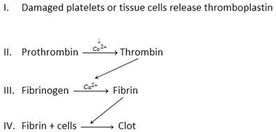
Blood coagulation is helped by thrombocytes.
Stage I is concerned with the formation of thromboplastin released from damaged tissue or platelets. Thromboplastin helps in the formation of the enzyme thrombokinase.
Stage II involves the conversion of prothrombin into thrombin with the help of thromokinase and calcium ions.
Stage III involves the conversion of a soluble protein fibrinogen in plasma to insoluble network of fibrous material called fibrin by the action of thrombin.
Stage IV is the formation of red solid mass called blood clot by trapping of blood cells particularly RBCs by fibrin network.
Coagulation factors
|
Factor |
Name |
Factor |
Name |
|
I |
Fibrinogen |
VIII |
Antihemophilic factor |
|
II |
Prothrombin |
IX |
Christmas factor or plasma thromboplastin component (PTC) |
|
III |
Thromboplastin |
X |
Stuart factor or Stuart-Prower factor |
|
IV |
Calcium- ions |
XI |
Plasma thromboplastin antecedent (PTA) |
|
V |
Proaccelerin (Labile factor) |
XII |
Hageman factor |
|
VI |
Hypothetical factor |
XIII |
Fibrin stabilizing factor (FSF) |
|
VII |
Serum prothrombin conversion accelerator (Stable factor) |
|
|
Functions of blood : On basis of the above account, the general functions of blood can be briefly enumerated as follows:
(1) Blood is the fluid medium which transports different materials between various parts.
(2) The leucocytes of blood play the important role of defense by inactivating and destroying harmful toxins and invaders like bacteria, viruses, fungi and animal parasites.
(3) Blood leucocytes phagocytes and destroy cell debris and inert foreign particles in blood and tissues. Thus, these act as “scavengers” to clean the body’s internal environment.
(4) Blood maintains the normal temperature of body. It prevents a sharp rise or fall in temperature which may be caused in any tissue due to abnormal rate of metabolism.
(5) By coagulating at an injury, and by stimulating repairing of damaged tissues, the blood helps in rapid healing of wounds and injuries.
(6) Blood helps in the maintenance of a proper internal environment in the body by regulating the amount of salts, acids, bases and water, etc. in the tissue fluids.
Lymph
Lymph can be defined as blood minus RBCs but more WBCs. Lymph is chiefly made of plasma plus leucocytes. Most important centre for the formation of lymph is interstitial space. Interstitial fluid, intercellular fluid, tissue fluid and lymph all are same in composition. Exchange of materials between blood and tissue fluid occurs through blood capillaries.
Functions of lymph : The basic function of lymph is to bring back, into the vascular circulation, the cell debris, large colloid particles and the part of the blood plasma that had diffused out from arterial capillaries into the tissue fluid but has failed to return back into venous capillaries. The white corpuscles of the lymph are the same as those of the blood and have the same functions of defense and of assistance in tissue repair and healing. In intestinal wall, lymph capillaries, called lacteals, are specially meant for absorption of fats.
Differences between blood and lymph
|
S. No. |
Blood |
Lymph |
|
1. |
Red corpuscles present. |
These are absent. |
|
2. |
White corpuscles fewer, neutrophils most numerous. |
White corpuscles more; lymphocytes most numerous. |
|
3. |
Soluble proteins more than insoluble proteins. |
Insoluble proteins more than soluble proteins. |
|
4. |
Amount of nutrients and \[{{O}_{2}}\] comparatively more. |
Amount of nutrients and \[{{O}_{2}}\] comparatively less. |
|
5. |
Amount of \[C{{O}_{2}}\] and metabolic wastes normal. |
Amount of these much more. |


You need to login to perform this action.
You will be redirected in
3 sec

