Rattus Rattus (The common House Rat)
Category : 11th Class
Systematic position
Phylum - Chordata
Subphylum - Vertebrata or Craniata
Class - Mammalia
Order - Rodentia
Family - Muridal
Genus - Rattus
Species - rattus (Black Rat)
They are the common house rats which are cosmopolitan in distribution and found all over the world. They are herbivorous, fossorial and nocturnal animals and undergo hibernation. They show sexual dimorphism. They are prolific breeders. Fertilization is internal. The time interval between fertilization and birth (gestation) is about 22 to 23 days. They are completely grown at six to eight months of age. The rat breeds more than four times in a year producing 6-8 young ones in each litter. Newly born young ones are blind, deaf and without hairs. The mother feeds the young ones on milk. Average age of a rat is 3 years.
External morphology
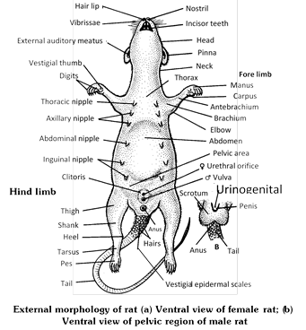
Their body is covered with hairs. The body is divisible into head, neck, trunk and tail.
Head : Head is broader posteriorly and tapers anteriorly as a naked terminal muzzle or snout. A pair of nostrils, shaped like inverted commas, is present above the mouth opening, which leads into nasal passages. Below the nostrils is the cleft upper lip, which exposes the two upper incisors. On the lateral sides of the head are large, paired bulging eyes. Eyelids have very fine and short eyelashes; the nictitating membrane is reduced. The head bears a pair of external ear or pinna at its posteriolateral position. The mouth is sub-terminal and located beneath the nostrils and remains guarded by upper and lower lips. Long, stiff, bristle-like hair known as pili lactiles or vibrissae are present on both sides of nostrils. They help the animal in measuring width of area through which the animal is to pass even in perfect darkness.
Neck : It is a short connective between head and trunk. With the help of neck the animal can bend its head in different directions.
Trunk : It is depressed fusiform major part of the body which has two parts-anterior narrow but stouter thorax and posterior wider softer abdomen. The ventral surface of female bears 6 pairs of teats or nipples, three pectoral (thoracic) and three inguinal (abdominal). The trunk bears two pairs of limbs, two forelimbs and two hindlimbs. Forelimbs are smaller than the hindlimbs. Each limb is made up of proximal segment (stylopodium), middle segment (Zeugopodium) and distal segment (autopodium). Five digits are present in autopodium of each limb. The first digit is thumb or pollex, which is much reduced with a peculiarly flattened nail and two phalanges. Nail is keratinized structure occupying position above the distal phalanx of each digit. Typical walking pads, the tori are present on the tips of digits, palm and at the base of palm. These are also present on the feet, but palms and soles do not have hair. Anus lies posteriorly at the base of tail.
Tail : It is quite long cylindrical and tapering structure that develops above the anus. It bears overlapping scales and sparse hair in between. Tail is used as a balancing organ.
Skin (Integument)
Histologically, the skin consists of outer epidermis and inner dermis.
(1) Epidermis : Epidermis is ectodermal in origin and is made up of stratified squamous epithelium. It consists of stratum germinativum (S. malpighi), S. spinosum, S. granulosum, S. lucidum and S. corneum. There are some layers of cells above the stratum lucidum which constitute the stratum corneum. The cells contain a protein called keratin and have lost all other internal structures including nuclei.
(2) Dermis : It develops from the mesoderm of embryo. It is composed of dense fibrous connective tissue with blood vessels , lymph vessels, nerve fibres, pigment cells etc.
Derivatives of skin : Hairs, cutaneous glands and claws are formed from the skin. Major skin glands are sudoriferous glands (sweat glands), sebaceous glands (oil glands), mammary glands (modified sweat glands), meibomian glands (modified oil glands, present along the edges of the eye lids) and ceruminous glands (wax glands, present in the external auditory canal of external ear).
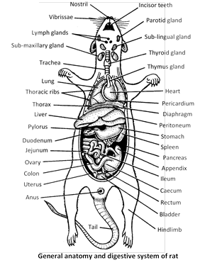
Digestive system
It consists of alimentary canal and digestive glands.
Alimentary canal : Alimentary canal is coiled tube of variable diameter. It begins at mouth and ends at anus. The various parts are mouth, buccopharyngeal cavity, oesophagus, stomach, small intestine (duodenum, jejunum and ileum), large intestine (caecum, colon and rectum) and anus.
(1) Mouth : The mouth opens in the buccal cavity that is surrounded by the vestibule, which is a space between the lips, cheeks and teeth.
(2) Buccopharyngeal cavity : It is space enclosed by two jaws. Buccopharyngeal cavity consists of broader buccal cavity in the anterior region and narrow pharynx in the posterior region. Jaws bear teeth. The teeth are heterodont, thecodont and monophyodont. Each jaw carries two incisors and six molars the incisors grow throughout life and act as growing teeth. A sharp cutting edge is maintained due to the absence of enamel on the surface. The canines and premolars are absent. A space called diastema occurs between incisors and molars. The dental formula is \[\frac{1003}{1003}\times 2=16\]. The middle of buccal cavity contains a muscular tongue. Taste buds occurs on tongue as well as lining of buccopharyngeal cavity. Unlike frog tongue is attaches posterioly. Behind lies pharynx. Pharynx is a common chamber for the passage of food and air.
(3) Oesophagus : It is a short tube situated dorsal to the trachea and it leads into the pear-shaped or somewhat semicircular stomach.
(4) Stomach : It is wide curved part of alimentary canal which lies on the left side behind the diaphragm. It has a greater curvature on left side, a lesser curvature on right side, cardiac orifice/valve where oesophagus open into it and pyloric sphincter is narrow posterior and where it meets duodenum. Stomach contain goblet cells for mucus, oxyntic cells for HCl and peptic cells for secretion of pepsinogen.
(5) Small intestine : Stomach leads into small intestine, which can be differentiated into three parts duodenum (U-shaped), jejunum (straight) and ileum (coiled). Digestive glands of small intestine secrete intestinal juice or succus entericus. The same contain lipase, nuclease, peptidase, lactase, sucrase and maltase enzymes.
(6) Large intestine : It has three part-caecum, colon and rectum. Caecum is slightly constricted about its middle. The constriction sub divides the caecum into two parts, the an apical and basal portion. The apical portion contains a distinct mass of lymphoid tissue forming the vermiform appendix. Caecum opens into the first part of large intestine, the colon which is divisible into an ascending, a transverse and a descending colon leads into rectum, which opens outside through the anus.
Digestive glands
(1) Salivary glands : There are three pairs of salivary glands.
(i) Sublingual glands
(ii) Submandibular glands and
(iii) Parotid glands.
Infraorbital salivary glands are reported but probably they are absent in rat, however, dogs have these glands.
(2) Liver : It is the largest gland of the body which is located in the upper and right side of the abdominal cavity below the diaphragm. The liver of rat consists of four lobes (left, middle, right and caudate) and the spigelian lobe is a part of caudate lobe. The cells of liver are called hepatocytes which secrete bile. Bile is carried to the duodenum by bile duct. Bile contains no digestive enzymes but helps in digestion of food in the small intestine. Gall bladder is absent in rat. Gall bladder is also absent in whale and horse.
(3) Pancreas : It is very diffused structure and is present between the duodenal loops. It secretes pancreatic juice which contain digestive enzymes such as trypsinogen (proenzyme), amylopsin and lipase. lslets of Langerhans of the pancrease secrete certain hormones such as insulin. Insulin converts glucose into glycogen in the liver and muscles.
(4) Gastric Glands : These are found in stomach and secrete gastric juice containing digestive enzymes (e.g., pepsin) and hydrochloric acid \[(HCl)\] which help in digestion of food.
(5) Intestinal Glands : These are present in the small intestine and secrete intestinal juice containing digestive enzymes (e.g., maltase, sucrase, lipase, etc.) which help in digestion of food.
Respiratory system
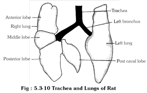
It consists of respiratory tract, two lungs and a mechanism for inspiration and expiration. Respiratory tract consists of nostrils, nasal chambers, internal nares, glottis, larynx, trachea, bronchi, bronchioles and alveoli. The nostrils lead into the olfactory or nasal chambers. The two nasal chambers lead into pharynx through internal nares. Pharynx contains a slit like glottis, which leads into voice box called larynx. Larynx passes into trachea and wind pipe which runs ventral to oesophagus. Trachea divides into two primary bronchi that pass into lungs. The lungs are placed one on an either side of the heart lie in the thoracic cavity and are covered by visceral pleura. There are three lobes of the right lung and only one in the left. Each lung possesses a large number of alveoli where gaseous exchange occurs between air and blood.
The circulatory system of rat consists of blood vascular system and lymphatic system.
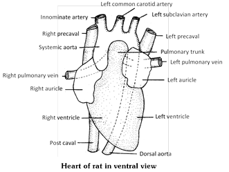
(1) Blood vascular system : Like other mammals rat possesses closed and double circulation. Blood vascular system comprises blood, heart and blood vessels.
Blood : The volume of blood is about 5-7 ml/100 gm body weight. The blood consists of blood plasma and three types of blood corpuscles namely RBCs (6-7 lac/cubic microlitre), WBCs (6-10 thousands/cubic microlitre) and platelets. RBCs are without nucleus on maturation. They contain haemoglobin (respiratory pigment). WBCs provide immunity and defence against diseases. The platelets help in clotting of blood.
Heart : The heart lies on the midline and placed obliquely in the thoracic cavity, surrounded by pericardial cavity. The heart has four chambers; the right atrium and right ventricle and the left atrium and left ventricle. Blood flows from the right atrium into the right ventricle via the tricuspid valve (right atrio-ventricular valve) with three cusps of fibrous tissue. Blood flows from the left atrium into the left ventricle via the bicuspid or mitral valve (left atrio-ventricular valve). Aortic and pulmonary valves each have three leaflets and called semilunar valve. The right cardiac arteries supply right and left atria, whereas the left cardiac arteries only supply to small portion of the left atrium. Well developed arterial and venous system similar to other mammals is found in the rat. Only the left aortic arch is present and two precavae are present in the rat. Hepatic portal system is present which comprises veins collecting blood from alimentary canal and supply to the liver after branching in capillaries. The renal portal system is absent.
(2) Lymphatic system : It consists of lymph, lymph vessels and Lymphatic nodes. Lymph is colourless fluid which is similar to blood but lacks red blood corpuscles and blood platelets. Lymph is formed by lymph capillaries from tissue fluid. Lymph capillaries join to form lymph vessels. At places lymph vessels bear lymph nodes. The latter contain minute channels where germs are entrapped by leucocytes. Lymph nodes also produce lymphocytes. Tonsils a type of lymphatic node are, however, absent. Lymph vessels form lymph ducts of two types, right and thoracic. They also open into veins.
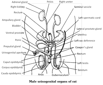
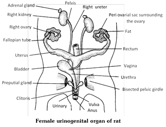
The excretory system includes paired kidney, ureters, a urinary bladder and urethra.
(1) Kidneys : There is a pair of kidneys which are dark red and bean shaped. The right kidney is slightly higher in position. The kidney consists of outer cortex and inner medulla. A kidney has numerous structural and functional units called nephrons (= uriniferous tubules). Each nephron is made up of Bowman’s capsule, proximal convoluted tubule (PCT), Henle’s loop and distal convoluted tubule (DCT). The Bowman’s capsule is a cup shaped structure which contains a meshwork of blood capillaries, the glomerulus. Filtration of metabolic wastes takes place in the glomerulus. Filtrate comes to the Bowman’s capsule from the glomerulus and then to the other parts of the nephron.
(2) Ureters : There is a pair of ureters. Each ureter arises from each kidney. Ureters carry urine from the kidneys to the urinary bladder.
(3) Urinary bladder : It is muscular sac-like structure in which two ureters open. The urinary bladder stores urine temporarily.
(4) Urethra : In male it carries both urine and semen. In female it carries urine only. Thus in male rat there is only one urinogenital aperture to pass urine and semen. However in female rat both urinary and genital apertures are separate.
Nervous system
The nervous system is divisible into three main parts :
(1) Central Nervous System (CNS) : It is a hollow, dorsally placed structure lying along the middorsal axis of the body. It comprises the brain and spinal cord. The brain is lodged in the skull while spinal cord is enclosed by the vertebral coloumn.
(2) Peripheral Nervous system (PNS) : The nerves arising from the central nervous system constitute the peripheral nervous system. The nerves which originate from the brain and spinal cord are known as cranial nerves and spinal nerves respectively.
(3) Autonomic Nervous System (ANS) : It controls and coordinates such organs which are under involuntary control. It consists of sympathetic and parasympathetic nervous system.
Brain : The brain is lodged in the cranial cavity (= cranium of the skull). It is covered by three membranes called meninges. The inner most membrane is called piamater, the next is the arachnoid mater (= archnoid membrane) and outer most is the duramater. The sub dural space is present below the dura mater and sub arachnoid space lies below the arachnoid mater. These spaces are filled with a fluid. The meninges are protective in function.
The brain is composed of two large hemispheres separated by a median fissure. The cerebral hemispheres form the largest part of the brains. The posterior portion of the brain is composed of medulla oblongata which tapers in the spinal cord. The spinal cord is a long tube like thick walled structures emerges out through the foramen magnum of the skull and passes through neural canal of vertebral column. The peripheral nervous system comprises the nerves arising from the brain and spinal cord.
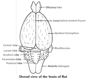

Cranial nerves : Rat has 12 pairs of cranial nerves originating from brain.
Spinal Nerves : 33 pairs which come out from intervertebral foramina. Spinal nerves are mixed. They supply various organs in the area of their origin.
Sense Organs
Skin has tangoreceptors (receptors of touch), thermoreceptors (receptors of temperature) algesioreceptor (receptors for pain) and rheoreceptors (receptors for current or vibrations). Gustatoreceptors (taste receptors) occur in the form of taste buds over tongue and posterior part of palate. Olfactoreceptors are located in olfactory epithelium present in nasal chambers. They perceive the sensation of smell. Smell is also perceived by a pair of Jacobson’s organs present in the wall of buccal cavity of Rat. Organs of sight are eye while statoacoustic organs are ears.
Male Reproductive system : The male reproductive organs of a rat are a pair of testes, epididymis, vasa deferens, urethra, penis and spermatic cord.
(1) Testes : A pair of testes is found in the scrotal sacs. Each testis is an elongated and ovoid body attached posteriorly to scrotal sac by gubernaculum. Testis of male rat descends in the scrotal sacs between the 30th to 40th day of life through inguinal canal. The inguinal canal remains open through out life, but during sexually inactive period, the tests may be withdrawn into abdominal cavity.
(2) Epididymis : These are paired structure. Each epididymis is a mass of long narrow coiled tubule lying along the testis which consists of anterior caput epididymis, middle corpus epididymis and posterior cauda epididymis. Epididymis stores the sperms.
(3) Vasa deferentia : There is a pair of vasa deferentia. A vas deferens arises from the cauda epididymis. Vasa deferentia carry sperms.
(4) Seminal vesicles : There is a pair of seminal vesicles which are large and lobulated except for the smooth tip which is doubled back upon itself. They are not store houses for sperms. Their secretion is alkaline and forms the bulk of seminal fluid (semen).
(5) Urethra : It is divided into three parts –
(i) Prostatic urethra is surrounded by the prostate gland.
(ii) Membranous urethra is the shortest portion and runs from the prostate to the bulb (base) of the penis.
(iii) Penile urethra passes through the penis and opens at the tip of the penis as urinogenital aperture.
(6) Penis : It is a copulatory organ which is covered by a loose sheath, the prepuce. The penis of the rat has a bony process called the os penis. Penis bone is also present in bat, dog, walrus and whale.
Accessory Glands : Male accessory sex glands –
(1) Ampullary glands : The outer end of the vas deference near the entrance into the urethra is enlarged into ampulla, which contain ampullary glands to secrete mucus.
(2) Vesicular glands : These are branched glands which originate from the vas deferens behind the ampulla.
(3) Coagulating glands : Closely applied along the minor curvature of the seminal vesicles and within the same sheath are the coagulating glands. The secretion of these glands serve to coagulate the seminal fluid (Semen).
(4) Prostate glands : There are two prostate glands whose secretion is rich in citric acid, lipid and acid phosphatase.
(5) Cowper’s glands : (Bulbo-urethral glands) : These are one pair which originate from the urethra at the base penis. They produce a secretion during sexual excitement which protects the sperms from traces of acids found in the urethra (as the urine also passes through the penile urethra).
(6) Preputial glands (Glands of Tyson) : They develop from the skin forming prepuce. They are modified sebaceous (oil) glands which secrete peculiar odorous secretion.
Female reproduction system : The female reproductive organs consists of a pair of ovaries, fallopian tubes, uteri, a single common vagina and a clitoris.
(1) Ovaries : Ovaries are paired small yellowish compact structure suspended in the body cavity by mesovarium.
(2) Fallopian Tubes (Oviducts or uterine tubes) : There is one pair of convoluted Fallopian tubes. Each Fallopian tube begins with fimbriated funnel which receives ova from the ovary. As the fertilization is internal, it takes place in the dilated upper most portion of the Fallopian tubes.
(3) Uterus (Womb) : The uterus is a hollow muscular structure. The uterine horns are fused near vagina. The wall of the uterus consists of outer covering of peritoneum, the perimetrium, middle layer of smooth muscle fibres, the myometrium and inner layer of simple columnar epithelium, the endometrium. The embryo gets attached to the uterine wall through placenta. Embryonic development takes place in the uterus. Placenta provides the physiological connection between developing foetus and uterine wall (endometrium) of the mother.
(4) Vagina : It is a tubular structure which extends from the uterus and opens outside as vaginal opening (= vulva). Penis of the male rat is inserted into the vagina during copulation. The vagina also helps to deliver the young ones at the time of birth.
(5) Clitoris : It corresponds to the penis of the male but it is reduced in size and does not have any passage (it is solid structure). The clitoris is found anterior to the vulva.
Accessory Glands
(1) Vestibular glands : These are small mucous glands which open on the surface of the vestibule of the vagina.
(2) Bulbo-urethral glands : These are small glands which are present in relation with the urethra.
(3) Preputial glands : There is one pair of large preputial glands which are near the tip of the clitoris.
(1) Rat is an important pest of crops, stored grains, fruits, vegetables, etc.
(2) Rat destroys field by making burrows and tunnels which often provide shelter to snakes.
(3) Rat makes burrows in the houses and causes damage to the household including books, clothes, food, etc.
(4) Rat is a host of rat flea, Xenopsylla, which is the vector for the disease bubonic plague.
(5) Rat is an important component of food chain as several animals like cats, snakes, mongoose, owls and some other birds use it as their food.
(6) The albino rat that is commonly used for teaching and researches in institutions is a product of laboratory breeding.
(7) The albino rats are also used as an experimental animal to test drugs that are to be finally used by the human beings.


You need to login to perform this action.
You will be redirected in
3 sec

