Epithelial Tissue
Category : 11th Class
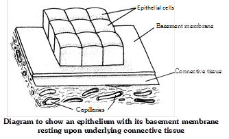
An epithelium is a tissue composed of one or more layers of cells that cover the body surface and lines its various cavities. It serves for protection, secretion and excretion. The word ‘epithelium’ (G. epi = upon, thele = nipple) was introduced by Ruysch. They are located on the outer surfaces of organs, including the skin. They form the linings of tracts, cavities and vessels. Epithelial tissue evolved first in animal kingdom. It originate from all the three primary germ layers.
Structure
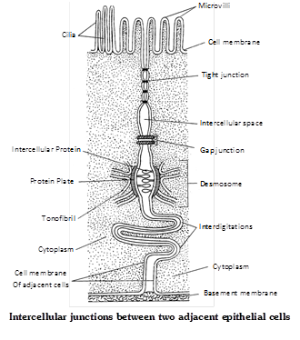
Cells are arranged in one or more layers, cells are compactly arranged and there is no inter cellular matrix between them. Neighbouring cells are held together by intercellular junctional complexes like desmosomes, tight junctions, interdigitations etc. The cells of lowermost layers always rest on a non living basement membrane or basal lamina. Basement membrane is made up of no cell product of epithelial tissue. It is formed of mucopolysaccharides, glycoprotein and collagen or reticular fibres. Blood vessels are absent in the epithelial tissues. However, nerve endings may penetrate the epithelium. It posses very high capacity of renewal (mitotic cell division). The following types of modifications and junctions are found in the plasma membrane of adjacent epithelial cells to keep the cells together.
Microvilli : It is simple and minute cytoplasmic processes arising from free exposed surfaces of the cell. They absorb material. e.g. Intestine.
Stereocilia : It is non-motile cytoplasmic processes. e.g. Epididymis, vas deference.
Kinocilia : It is contractile motile fibrous processes arising from basal granules. e.g. Oviduct, Fallopian tube.
Tight junctions (Zona occludens) : At certain places the plasma membranes of adjacent cells are tightly packed or even fused together. e.g. Brain.
Desmosomes : Desmosome is present in epithelial tissue. They consist of thickened area and several fine tonofibrils extending from each plasma membrane into cytoplasm of respective cells. Macula adherens is a kind of desmosome. e.g. Vagina, Urinary bladder.
Gap junction : At place, the adjacent cells form ion-rich gap junctions for intercellular communication and chemical exchange. These junctions probably do not provide physical support.
Interdigitations : These are interwoven finger-like processes of plasma membranes of adjacent cells.
Intercellular bridges : These are minute projections that arise from adjacent cell membranes. The intercellular bridges make contact with one another.
Functions
Epithelial tissues have a wide spread distribution throughout the body and serve several important functions –
(1) Generalized protection is the most important function of membranous epithelium. It is the relatively tough and impermeable epithelial covering of the skin that protects the body from mechanical and chemical injury and also from invading bacteria and other disease causing micro-organisms.
(2) Epithelial structures specialized for sensory functions are found in the skin, nose, eye and ear.
(3) Glandular epithelium is specialized for secretory activity, secretory products include hormones, mucous, digestive juices and sweat.
(4) The epithelium lining of the gut and respiratory tracts allows the absorption of nutrients from the gut.
(5) It is the specialized epithelial lining of kidney tubules that makes the excretion and concentration of excretory products in the urine.
(6) Ciliated epithelium moves fluid, mucous and other materials in the organs it lines.
(7) Germinal epithelium of the seminiferous tubules and ovaries produces spermatozoa and ova respectively.
(8) The ability of epithelia to regenerate quickly helps in the healing of wounds.
(9) Pigmented epithelium of retina darkens the cavity of eyeball.
(10) The epithelia check the absorption of harmful or unnecessary materials.
(11) Epithelium of alveoli of the lungs brings about exchange of gases between blood and air.
(12) Epithelium also produce exoskeletal structures such as scales, feathers, hair, nail, claws, horns and hoofs.
Types of epithelial tissue
Mainly based on the location and functions of tissue it is following types –
(1) Simple epithelium : It is simple in structure and basically formed by single layer cells.
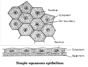
(i) Simple squamous epithelium : It is consists of only one layer of flat, scale like cells, usually polygonal cells which are closely fitted together like the tiles of a mosaic. It is also known as pavement epithelium. e.g., It forms lining of blood vessels, lymph vessel, heart, peritoneum, pleura, Bowman’s capsule, thin segment of loop of Henle and lung alveoli.
(ii) Simple cuboidal epithelium : The simple cuboidal epithelium is composed of one layer of cuboidal shaped cells resting on a basement membrane. The nuclei are situated centrally. e.g. the cuboidal epithelium is present in the small salivary and pancreatic ducts, thyroid vesicles, parts of membranous labyrinth, PCT, DCT, ovaries, seminiferous tubules of testes, ciliary bodies, choroid, iris of eyes, thin bronchioles and sweat gland of mammalian skin.

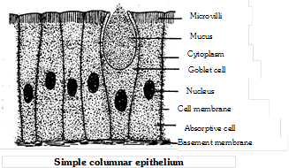
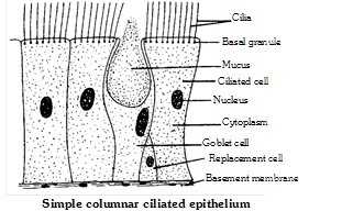
(iii) Simple columnar epithelium : It consists of a single layer cells, many of which have modified structure. Three common modifications are goblet, cilia and microvilli. Simple columnar epithelium is present in the stomach and intestine. e.g. located inner lining of gall bladder and bile duct. It also occurs in the gastric gland, intestinal glands, pancreatic lobules.
(iv) Simple ciliated epithelium : It bears numerous delicate hair like outgrowths called cilia arising from basal granules help to create a current to transport the materials. The ciliated epithelium is of two types :
(a) Ciliated columnar epithelium : It lines respiratory tract (Lower end of bronchi), fallopian tubes (oviducts), ventricles of brain (ependyma), central canal of spinal cord, tympanic cavity.
(b) Ciliated cuboidal epithelium : It occurs in certain parts of nephrons of the kidneys.
(v) Pseudostratified epithelium : It is always consist of single layer of irregularly shaped columnar cells, touches the basement membrane. The long cells have oval nuclei however,
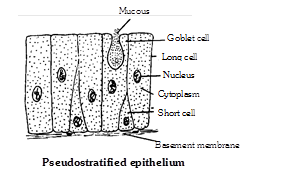
Short cells have rounded nuclei although epithelium is one cells thick, but it gives the appearance of a stratified epithelium, hence it is called pseudostratified epithelium. Mucous secreting goblet cells are numerous and cilia are present. It is of two types –
(a) Pseudostratified columnar ciliated epithelium : It is found in the lining of trachea and bronchi (Upper).
(b) Pseudostratified columnar epithelium : It is found in certain segments of human male urethra and parotid salivary gland, vasa deferentia and epididymis.
(c) Stratified squamous epithelium : The cells in the deepest layer are columnar or cuboidal with oval nuclei. It is called germinative layer. The cells of this layer divide by mitosis to form new cells.
(2) Compound epithelium : It is complexed in structure and basically formed by two or more than two layers of cells.
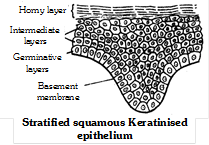
(i) Stratified squamous keratinised epithelium : Stratified squamous epithelium is characterized by multiple layers of cells with typical flattened squamous cells at the free or outer surface of the sheet. The presence of keratin in these cells contributes to the protective qualities of skin covering the body surface. Keratin is dead and waterproof so it protects the underlying tissues from abrasion and infection e.g. epidermis of the skin of land vertebrates.
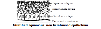
(ii) Stratified squamous non keratinised epithelium : Its free surface is moist, and the outer epithelial cells, unlike those found in the skin, do not contain keratin. This type of epithelium serves a protective function. It is found lining the oral cavity (buccal cavity), pharynx, oesophagus, anal canal, lowerpart of urethra, vocal cords, vagina, cervix (lower part of uterus) and cornea of eyes.
(iii) Stratified cuboidal epithelium : It is consists of two or more rows of low cuboidal-shaped cells which are arranged randomly over a basement membrane. It is found in the sweat gland ducts, larger salivary and pancreatic ducts.
(iv) Stratified columnar epithelium : It is protective epithelium has multiple layers of columnar cells, only the most superficial cells are truly columnar in appearance. Epithelium of this type is rare. It is found in male urethra and in the mucous layer near the anus. It also lines mammary gland ducts and epiglottis.

(v) Stratified columnar ciliated epithelium : It lines the larynx and upper part of the soft palate.
(3) Specialized epithelium : This type of epithelium are specialized to perform specific activity hence, specialized in structure also. They are as follows –
(i) Transitional epithelium (Urothelium) : It is often consists ten or more layers thick. It lacks germinative layer, basement membrane. Stratified transitional epithelium is typically found in the body areas such as the wall of urinary bladder, ureter and renal pelvis. It is located in all the hollow viscera subjected to stress and protects organ wall from tearing.
(ii) Neurosensory epithelium : Olfactory mucosa, called Schneiderian membrane, lining of internal nares, retina of eyes and epithelial covering of tongue containing taste buds are examples of neurosensory epithelia. The sensory cells bear, at their free ends, slender “sensory hairs” to receive specific stimuli. Basely, these cells are connected, by means of synapses, with fine fibrils of sensory nerves.
(iii) Pigmented epithelium : The epithelial cells of the basal layer of retina contain pigment. Hence, this layer is often referred to as a pigmented epithelium. e.g. – Pigmented layer of retina, iris and skin.
(iv) Germinal epithelium : Specialized cuboidal cells capable of producing gametes as found in gonads. Germinal epithelium produces gametes e.g., ova (Female gametes) and sperms (Male gametes)


You need to login to perform this action.
You will be redirected in
3 sec

