Answer:
The diagram representation of
amicrosporangium is shown below
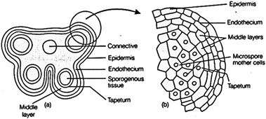
 (a) Transverse
section of a young anther (b) Enlarged view of one microsporangium showing wall
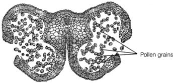
layers (c) Mature dehisced anther showing pollen grain
In a transverse section, a
typicalmicrosporangium is circular in outline and is surrounded byfour wall
layers.
(a)
Epidermis The epidermis is the outermost protective layer. It is composed
oftangentially flattened cells. The cells are closely fitted and have thick
walls which ishelpful in the dehiscence of anther.
(b)
EndotheciumIt is present below the epidermis and expands radically with
fibrous'thickenings, at maturity these cells loose water, at contract and help
in dehiscence ofpollen sac.
(c) Wall Layersit is present
between well marked endothecium and tapetum. These arethin walled layers,
arranged in one to five layers, which also help in dehiscence ofanther.
(d) TapetumIt is the
innermost wall layer with large cells, thin cell walls, abundantcytoplasm and
have more than one nuclei. Tapetum is a nutritive tissue whichnourishes the
developing pollen grains.The centre of the microsporangium consists of
sporogenous tissue, which undergoes meiotic divisions to form microspore
tetrads. This process is known asmicrosporogenesis.
(a) Transverse
section of a young anther (b) Enlarged view of one microsporangium showing wall
layers (c) Mature dehisced anther showing pollen grain
In a transverse section, a
typicalmicrosporangium is circular in outline and is surrounded byfour wall
layers.
(a)
Epidermis The epidermis is the outermost protective layer. It is composed
oftangentially flattened cells. The cells are closely fitted and have thick
walls which ishelpful in the dehiscence of anther.
(b)
EndotheciumIt is present below the epidermis and expands radically with
fibrous'thickenings, at maturity these cells loose water, at contract and help
in dehiscence ofpollen sac.
(c) Wall Layersit is present
between well marked endothecium and tapetum. These arethin walled layers,
arranged in one to five layers, which also help in dehiscence ofanther.
(d) TapetumIt is the
innermost wall layer with large cells, thin cell walls, abundantcytoplasm and
have more than one nuclei. Tapetum is a nutritive tissue whichnourishes the
developing pollen grains.The centre of the microsporangium consists of
sporogenous tissue, which undergoes meiotic divisions to form microspore
tetrads. This process is known asmicrosporogenesis.


You need to login to perform this action.
You will be redirected in
3 sec

