Answer:
(a) Structure of Brain The human brain is well
protected by the skull. The brain can be divided into three major parts
forebrain, midbrain and hindbrain.
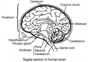 (i) Forebrain The various parts of forebrain are
cerebrum, thalamus and hypothalamus. Cerebrum is responsible for complex
functions like intersensory associations, memory and communication.
(ii) Midbrain The midbrain is located
between the thalamus/hypothalamus of the forebrain and pons of the hindbrain.
(iii) Hindbrain The hindbrain comprises pons,
cerebellum and medulla (also called the medulla oblongata). The medulla
contains centres, which control- respiration cardiovascular reflexes and
gastric secretions.
(b) Structure of Eye The wall of the human eye ball
is composed of three layers. The external layer is called the sclera. The
anterior portion of this layer is called the cornea. The middle layer, choroid,
contains many blood vessels and looks bluish in colour. The ciliary body itself
continues forward to form a pigmented and opaque structure called the iris. The
eye ball contains a transparent crystalline lens. In front of the lens, the
aperture surrounded by the iris is called the pupil.
(i) Forebrain The various parts of forebrain are
cerebrum, thalamus and hypothalamus. Cerebrum is responsible for complex
functions like intersensory associations, memory and communication.
(ii) Midbrain The midbrain is located
between the thalamus/hypothalamus of the forebrain and pons of the hindbrain.
(iii) Hindbrain The hindbrain comprises pons,
cerebellum and medulla (also called the medulla oblongata). The medulla
contains centres, which control- respiration cardiovascular reflexes and
gastric secretions.
(b) Structure of Eye The wall of the human eye ball
is composed of three layers. The external layer is called the sclera. The
anterior portion of this layer is called the cornea. The middle layer, choroid,
contains many blood vessels and looks bluish in colour. The ciliary body itself
continues forward to form a pigmented and opaque structure called the iris. The
eye ball contains a transparent crystalline lens. In front of the lens, the
aperture surrounded by the iris is called the pupil.
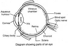 There
are two types of photoreceptor cells namely rods and cones. The optic nerves
leave the eye and the retinal blood vessels enter it at a point medial to and
slightly above the posterior pole of the eye ball. Photoreceptor cells are not
present in that region and hence, it is called the blind spot.
(c) Structure of Ear Anatomically, the ear can be
divided into three major sections called the outer ear, the middle ear and the
inner ear.
(i) Outer ear The outer ear consists of the pinna
and external auditory meatus (canal).
(ii) Middle ear The middle ear contains three ossicles
called malleus, incus and stapes which are attached to one another like a
chain. The malleus is attached to the tympanic membrane and the stapes is
attached to the oval window of the cochlea. An eustachian tube connects the
middle ear cavity with the pharynx.
(iii) Inner ear The fluid-filled inner ear called
labyrinth consists of two parts, the bony and the membranous labyrinths.
There
are two types of photoreceptor cells namely rods and cones. The optic nerves
leave the eye and the retinal blood vessels enter it at a point medial to and
slightly above the posterior pole of the eye ball. Photoreceptor cells are not
present in that region and hence, it is called the blind spot.
(c) Structure of Ear Anatomically, the ear can be
divided into three major sections called the outer ear, the middle ear and the
inner ear.
(i) Outer ear The outer ear consists of the pinna
and external auditory meatus (canal).
(ii) Middle ear The middle ear contains three ossicles
called malleus, incus and stapes which are attached to one another like a
chain. The malleus is attached to the tympanic membrane and the stapes is
attached to the oval window of the cochlea. An eustachian tube connects the
middle ear cavity with the pharynx.
(iii) Inner ear The fluid-filled inner ear called
labyrinth consists of two parts, the bony and the membranous labyrinths.
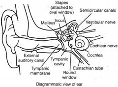 The
organ of Corti is a structure located on the basilar membrane which contains
hair cells that act as auditory receptors. The inner ear also contains a
complex system called vestibular apparatus, located above the cochlea. The
vestibular apparatus is composed of three semi-circular canals and the otolith
organ.
The
organ of Corti is a structure located on the basilar membrane which contains
hair cells that act as auditory receptors. The inner ear also contains a
complex system called vestibular apparatus, located above the cochlea. The
vestibular apparatus is composed of three semi-circular canals and the otolith
organ.


You need to login to perform this action.
You will be redirected in
3 sec

