Male Reproductive System
Category : 12th Class
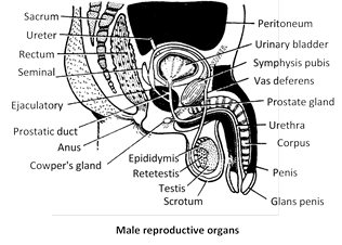
The male reproductive system consists of a scrotum, a pair of testes, vasa efferentia, a pair of epididymis, a pair of vasa deferentia, a pair of ejaculatory ducts, a urethra, a penis and certain accessory sex glands.
Reproductive organs
(1) Scrotum : The scrotum is a pouch of pigmented skin arising from the lower abdominal wall and hanging between the legs. The testes originate in the abdominal but latter, during the seventh month of development, descend permanently into the respective scrotal sac through passages termed inguinal canal.
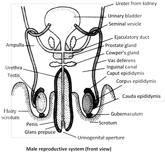
A spermatic cord connects testis with abdominal cavity. It consists of connective tissue that encloses an artery, a vein, a lymph vessel, a nerve, cremaster muscle and a vas deferens. A testis rests in it chamber over pad called gubernaculum.
The scrotal sac of male homologous to female's labia majora.
(2) Human Testes : The testes are the primary sex organs. They are about \[45\,\,cm\] long, 2.5 cm wide and 3 cm thick. They are suspended in the scrotal sacs by spermatic cords. Each testes weights about \[10-15\text{ }gms.\]
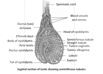
Each testis has three coverings - tunica vaginalis, tunica albuginea and tunica vasculosa.
In growth of the tunica albuginea, called septa, divide the testis into some 200 to 300 lobules. Each testicular lobule contains \[13\] highly convoluted seminiferous tubules, blood vessels and nerve embedded in loose connective tissue. A total of about 750 seminiferous tubules occur in each testis.
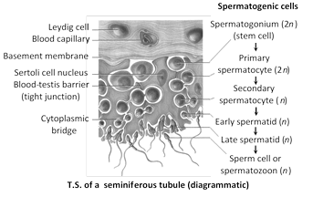
Each seminiferous tubules is lined by germinal epithelium, seminiferous tubules is the site of spermatogenesis. The process occurs in waves along the length of the tubule, taking about 9 weeks (63 days) to complete in man. Seminiferous tubules contain 3 types of cells -
(i) Germ cells : Germ cells or primordial germ cells arise from yolk sac endoderm and enter the testes early in development. These are spermatogenic cells, by mitotic divisions, produce spermatogonia into the lumen of the seminiferous tubule. The spermatogonia grow into primary spermatocytes which undergo meiosis, producing haploid cells, first secondary spermatocytes and then spermatids. Spermatids differentiate by a process of spermiogenesis into dimorphic haploid sperm (containing X or Y chromosome). Mature spermatozoa lie free in the cavity of the seminiferous tubules.
(ii) Somatic cells / Sertoli cells / Sustentacular cells / Nurse cells : These are supportive nutritive and secrete a polypeptide hormone called inhibin and a steroid estradiol which interferes with spermatogenic activity and kinetics of sperm production.
(iii) Leydig cells ( = Interstitial cell) : Leyding cells endocrine cell of testes which lie in the form of clusters or singly in the interstitium (=space between seminiferous tubules).
These are secrete a sex steroids called androgen by using cholesterol. The cells contain a rich repertoire of enzymes which facilitate formation of pathways for steroid biosynthesis and biotransformation. These enzymes are called steroid-dehydrogenases.
The seminiferous tubules open into rete testis.
(3) Vasa efferentia : Rete testis is connected to caput epididymis by \[1220\] fine tubules called vasa efferentia or ductuli efferentes. Their lining epithelium is ciliated for conducting sperms.
Tubuli recti, rete testis and ductuli efferents constitutes an intertesticular genital duct system. The cells of vasa efferens are columnar ciliated.
(4) Epididymis : From rete testis sperms moves into a series of coiled efferent ducts in epididymis that empty into a single tube called ductus epididymis present inside epididymis as highly coiled tube, measures about 6 m (20 ft) in length. It is lined by pseudostratified columnar epithelia.
(i) Upper part (Heads) : Caput epididymis or globus major.
(ii) Middle part : Corpus epididymis or globus normal.
(iii) Basal part (Tail) : Cauda epididymis or globus minor.
In epididymis the sperms are stored for a few hours to a few days till sent out through ejaculation.
The epididymis shows peristaltic and segmenting contraction at intervals to push the spermatozoa away from the testis.
Testis and epididymis are together called testicle.
(5) Vasa deferentia (Singular-vas deferens) : The vas deferens is a continuation of the cauda epididymis. It is about 45cm. long and is slightly coiled at first but becomes straight as it enters the abdominal cavity through the inguinal canal.
Vasa deferentia (ducti deferentes) conduct sperms from epididymis to urethra and lined by pseudostratified columnar epithelia.
Surgical interference (vasectomy) of vas deferens ensure successful non-reversible male contraception.
Difference between Vasa efferentia and Vasa deferentia
|
S.No. |
Vasa efferentia |
Vasa deferentia |
|
1. |
Arise from the rete testes. |
Arise from the cauda epididymis. |
|
2. |
Vary from 15 to 20 in number. |
Are only 2 in number. |
|
3. |
Are fine and convoluteds |
Are thick slightly coiled in the scrotum, straight in the abdomen |
|
4. |
Lining bears many ciliated cells. |
Lining has sterocilia on many cells. |
|
5. |
Carry spermatozoa from rete testes to caput epididymis |
Carry spermatozoa from cauda epididymis to ejaculatory ducts. |
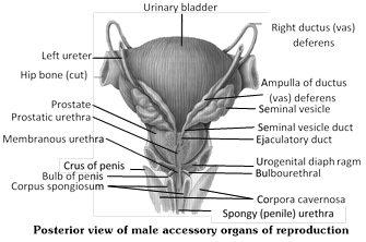
(6) Ejaculatory ducts : They are short (2 cm) straight muscular tubes each formed by union of a vas deferens and duct of seminal vesicle where ejaculate is formed by mixing of sperms with secretion of seminal vesicle. The two ejaculatory ducts join the urethra within prostate gland.
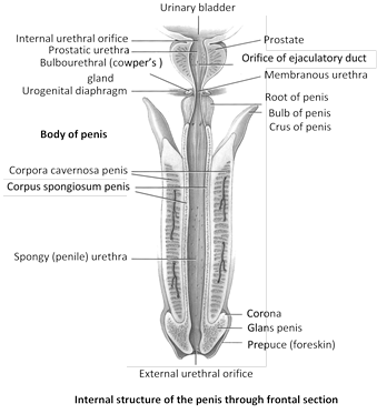
(7) Urethra : It is the urinary duct leading from the bladder. Urethra passes through prostate gland, urinogenital diaphragm, and penis. From the point it is joined by ejaculatory ducts, it carries urine as well as spermatozoa and secretions of the seminal vesicles. It also receives secretion of the prostate and cowper's glands. Urethra is some 20 cm long and passes through the penis. The urethra has 4 regions –
(i) Urinary urethra : It carries only urine.
(ii) Prostatic urethra : It is a short proximal part which is surrounded by prostate gland.
(iii) Membranous urethra : It is a short middle part, without any covering, is smallest part of urethra.
(iv) Penile urethra : It is a long distal part that passes through the penis, also known as spongy uethra.
The penile part is also called spongiose urethra because it lies inside corpus spongiosum.
(8) Penis : The penis is an erectile copulatory organ. It consist of a long shaft that enlarges to form an expanded tip, the glans penis. It is covered by a loose, retractable fold of skin, the prepuce or foreskin. Under the skin, the penis contains three columns of erectile tissue : two cylinders of the corpora cavernosa of the penis, placed dorsally, and one cylinder, the corpus spongiosum, along the ventral side. The corpora cavernosa of the penis and the urethra are covered by dense connective tissue, the tunica albuginea. Both urine and semen are carried out of the body through the penis. Corpus spongiosum contains the spongy urethra. Margins of glands penis known as corona.
The penis of opposum, bandicoot etc. is doubled branched.
Accessory sex glands : The substances secreted by the accessory, sex glands help in reproduction these are –
(1) Seminal vesicles : The seminal vesicles are long pouches with muscular wall; they secrete spermatozoa activating substances, such as fructose, citrate, inositol, prostaglandins and several proteins, sperms use fructose as a respiratory substrate. Seminal fluid maintains viability and motility of sperms.
Seminal vesicle secretes a alkaline, nutritive fluid which forms main part i.e., 60 % of the semen. It is also called uterus-masculinus. It forms from the mullerian duct of the embryo. In females, these ducts form the ovi-ducts. The seminal vesicle do not store sperms. Seminal vesicles are found between urinary bladder and rectum.
(2) Prostate gland : The prostate gland surrounds the first portion of the urethra. This gland secretes an slightly acidic fluid (pH about 6.5) which forms 25% part of the semen. The secretion nourish and activates the spermatozoa to swim. It is essential for sperm motility (removal causes sterlity).
In the secretion of prostate–gland citric acid, calcium and phosphate, Fibrinogen and Fibrinolysin is present. The secretion of the prostate gland combines with the secretion of seminal vesicle and so the semen gets coagulated. In the coagulated semen, the mobility of sperms is reduced and so their energy is conserved. After sometime due to fibrinolysins, semen again liquefies and in this semen now the sperms can move.
(3) Cowper's glands : These are also termed as Bulbourethral glands. 1st pair of Cowper's glands are attached to urethra. They secrete alkaline mucus which is discharged into the spongy part of urethra. The mucus lubricates the reproductive tract. This serves to neutralize any acid of urine remaining in the urethra. Secretion of Cowper's glands is produced before the ejaculation of semen.
Secretion of Cowper's glands carries some spermatozoa released before ejaculation. This is one of the reasons for the high failure rate of the withdrawal method of birth control.
(4) Perineal or Rectal glands : These are found both in males and females during the breeding season, these glands secrete and odoriferous liquid which has pheromones or Ectohormones in it. Its smell attracts the animal of opposite sex, found in herbivorous and carnivorous mammals.
In man, Perineal or Rectal glands are absent.
(5) Other glands : Prepuce contains preputial glands which produce a sebaceous substance which together with desquamated epidermal cells forms a whitish, pasty, foul-smelling accumulation, called smegma, about the base of the glans penis beneath the prepuce.
Semen : The products of the testes (spermatozoa) and prostate gland, alongwith fluid from the seminal vesicle, are collectively knows as semen. It is a milky, viscus and alkaline \[(pH\,\,7.27.7)\] fluid ejaculated by male reproductive system during orgasm. The volume of ejaculate varies from person to person. Abstinence play a role in this. Each ejaculate measures 3.5 ml and contains \[50150\] million sperm/ml i.e. 250 million \[\text{ }525\] million (average \[\text{ }400\]million).
The life span of human sperm after ejaculation is \[2448\text{ }hrs.\]Crayopreservation enhances the longevity of sperm. The rate of active moment of sperm is \[1.53.0\text{ }mm\]per minute in uterine endometrium.
Semen has chemicals for nourishing the sperms (e.g., - fructose), neutralizing the acidity of urethra and vagina (e.g., - bicarbonate), stimulating movements in female tract (e.g., - prostaglandins). pH of semen \[\text{ }7.27.7.\]
A person with a sperm count below 20 million will be physiologically sterlile. Fusion of defective sperm (e.g.,\[22+xy\]) with ovum causes many birth defects e.g., klinefelter's syndrome.
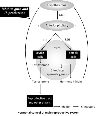
Hormonal control of male reproductive system : The growth, maintenance and functions of secondary sex organs (epididymis, vasa deferentia, accessory glands and penis) are under the control of testosterone hormone secreted by Leydig's cells of testis, while those of seminiferous tubules and Leydig's cells are controlled by Follicular Stimulating Hormone (FSH) and Interstitial Cells Stimulating Hormone (ICSH) of anterior pituitary lobe respectively.
Onset of puberty in the male : Puberty is the period when reproductive organs become functional. It is triggered by the secretion of the hormone testosterone in the testes. This hormone brings about growth and maturation of the secondary sex organs and development of the accessory sex characters. The latter induce :
(1) Enlargement of the penis and scrotum.
(2) Broadening of the shoulders.
(3) Growth of body and facial hairs.
(4) Deepening of the voice duce enlargement of layrnx and thickening of vocal-cords.
(5) Increased development of musculature and bones.
(6) Increase in height so characteristic of male puberty.
Male sex act
The male sex act involves 3 phases :
(1) Erection : Erection of the penis is caused by rush of arterial blood into the empty sinuses of its spongy tissue on sexual excitement. As the spongy tissue distends, it compress the veins, inhibiting the flow of blood out of the tissue. Filling of tissue with blood is called vasocongenstion.
(2) Copulation : Mucus from the urethral glands, Cowper's glands and vaginal glands provides lubrication for copulation. Friction due to rhythmic movements of sexual intercourse stimulate the sensory cells of the glans penis. This stimulation releases semen into the proximal part of urethra by contraction of reproductive glands and ducts. This process is called emission. Then the rhythmic, wavelike contractions of the muscles at the base of the penis cause forceful discharge, called ejaculation, of semen into the vagina. One ejaculate (about 3 ml.) contains 200 to 400 million spermatozoa. Ejaculation marks the climax of copulation.
Orgasm : At the peak of sexual stimulation, pleasurable sensation, called orgasm. It occurs usually last only a few seconds.
(3) Subsidence of erection : After ejaculation, the arterioles to the penis contract, reducing the blood flow to the penis, and erection subsides. This often takes a few minutes.
Disorders of male reproductive system
Only a few are mentioned.
(1) Prostatomegaly (Prostatic hypertrophy) : This is enlargement of prostate gland. If often occurs in old age. The enlarged gland may block the urethra, causing frequent night urination (nocturia) or difficult or painful micturition. Prostate cancer is very common in men. It is treated surgically or with drugs.
(2) Impotence : This is inability of the male to achieve and or maintain erection of the penis long enough to engage in or complete copulation.
(3) Sterility : Inability of the male's sperm to fertilized the ovum, it may or may not be associated with impotence. Sterility also results from immobility and morphological abnormality of the sperms, and from low sperm count in the semen.


You need to login to perform this action.
You will be redirected in
3 sec

