Lymphatic System
Category : 11th Class
It is a part of greater circulation which begins in the tissue fluid with lymphatic capillaries which are always terminally closed. This system terminates into venous system near heart. The main components of this system are :
(1) Lymph : Lymph can be defined as blood minus RBC's. In addition to the blood vascular system all vertebrate possess a lymphatic system. It is colourless or yellowish fluid present in the lymph vessels. It is a mobile connective tissue like blood and is formed by the filtration of blood. This process involves the diffusion of substances from blood capillaries into the interstitial space which is, thus, the primary site of lymph formation. Two forces bring about a steady filtration of plasma fluid into the tissue spaces : capillary pressure \[(30-35\,\,mm\text{ }Hg)\] and colloid osmotic pressure in tissue fluid (8 mm Hg). After absorption by veins, a small amount of \[C{{O}_{2}}\] and waste material still remains in the tissue fluid which is absorbed in the lymphatic capillaries as lymph. So, we can say that lymph is modified tissue fluid.
Differences between lymph and blood
|
S.No. |
Characters |
Blood |
Lymph |
|
(1) |
RBC |
Present |
Absent |
|
(2) |
Blood platelets |
Present |
Absent |
|
(3) |
WBC |
Persent, generally 7000/cu mm |
Persent, generally 500-75000/cu mm |
|
(4) |
Plasma |
Present |
Present |
|
(5) |
Albumin : globulin |
Albumin>Globulin |
Albumin>Globulin |
|
(6) |
Fibrinogen |
More |
Less |
|
(7) |
Coagulation property |
More |
Less |
|
(8) |
Direction of flow |
Two way, heart to tissues and tissues to heart |
One way, tissues to heart |
|
(9) |
Rate of flow |
Fast |
Slow |
|
(10) |
Glucose, urea and \[C{{O}_{2}}\] |
Less |
More |
Hence, lymph can be represented as :
Lymph = Blood – [RBC + platelets + plasma proteins of high molecular weight]
Composition of lymph : Microscopic examination of lymph depicts that is contains a large number of leucocytes (mostly lymphocytes) ranging from 500 to 75,000 per cubic mm. No blood platelets present. The composition of the non cellular part of lymph (fasting) is as follows :
(i) Water 94% (ii) Solids 6%
(a) Proteins : Protein content is roughly half of the plasma and varies from 2.0 - 4.5%. It varies according to the part of the body from which is collected, i.e. in liver 6%, in limb 2% of intestinal part 4%. The varieties of proteins are found - albumin, globulin and fibrinogen. In addition to this, traces of prothrombin, fibrinogen.
(b) Fats : In fasting condition fat content is low but after a fatty diet it may be 5.0 - 15%.
(c) Carbohydrates : Sugar, 132.2 mgm per 100 ml.
(d) Other constituents : Urea, creatinine, chlorides, phosphorus, calcium, enzymes and antibodies (120 ml./hour).
(2) Lymphatic organs : In human primary lymphatic (lymphoid) organs of the body are the Red bone marrow and Thymus gland. They are called primary lymphatic organs because they produce B and T cells the lymphocytes that carry out immune response. Haemopoietic stem cells in red bone marrow gives rise to B Cell and pre-T cells. Pre-T cells then migrate to thymus gland. Secondary lymphatic organs are the lymph nodes and spleen.
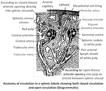
(i) Spleen : Spleen is mesodermal in origin. Spleen is the largest solid mass of reticulo-endothelial tissue in the body. In human measures about 12 cm (5 inch) in length and is situated in the left hypochondriac regions between the stomach and diaphragm. Like lymph nodes, spleen has hilus, where splenic artery, vein and efferent lymphatic vessels pass through. Spleen never filter lymph, because has no afferent lymphatic vessel. Histologically it is formed by following structure -
(a) Capsule : It is the outer covering of spleen formed of dense connective tissue and smooth muscles. The outer layer of the capsule is the serous coat formed of visceral peritoneum.
(b) Trabeculae : Narrow fold like septa or trabeculae extend inwards from the capsule, dividing the spleen tissue into several incomplete lobules.
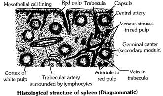
They provide support and convey blood vessels in to the interior of spleen.
(c) Splenic pulp : The reticulo-endothelial tissue is called splenic pulp. It contains a denser network of blood capillaries, small sinuses and fine blood vessels. The meshes of this network are studded with numerous splenic cells, red, blood corpuscles, macrophages and lymphocytes. The splenic pulp is of two distinct types -
(i) White pulp (ii) Red pulp
In mammal embryos the red pulp contains myelocytes, erythroblast and also megakaryocytes. These types of cells are not present in adult spleen except in certain pathological condition.
Function : Although located close to the alimentary canal, the spleen has nothing to do with digestive system. It is, in fact, an important constituent of the reticuloendothelial system of body and performs the following functions:
(a) Its macrophages engulf (= phagocytize) and destroy wornout blood corpuscles (RBC + platelets), dead and live pathogens, cell debris, pigment granules and other useless particulate materials, thus regularly cleaning the blood of its impurities.
(b) It is active haemopoietic organ. In foetal life, the red pulp possess myeloblast, erythroblast and megakaryocytes. Hence, in foetus, it produces blood. In adults, the red pulp possess macrophages, plasma cells and lymphocytes. So, in adults, it is not producing blood rather it is screening blood.
(c) In adults, it also serves as a sort of “blood bank”. Its sinuses act as “reservoirs of blood”.
(d) White pulp of spleen functions in immunity as a site of B cell proliferation into antibody-producing plasma cells.
(e) Spleen also acts as Graveyard or Slaughter house of worn out RBCs.
(f) Haemolysin is formed in spleen (Lysolecithin).
(g) Haemoglobin is broken down into haem and globin by spleen.
Besides all these functions, the primary function of spleen is that it assists liver and helps in maintaining the composition of blood.
(ii) Thymus gland : In human thymus is located in mediastinum, between the lungs. The two thymic lobes devide into lobules by trabeculae. Each lobule consist of cortex and medulla. Cortex composed of tightly packed lymphocytes, epithelial cells, Macrophages. Pre-T cells migrats (via blood) from red bone marrow to thymus, where they proliferats and develop into mature T cells. Medulla consist of mostly of epithelial cells and more widely scattered lymphocytes. Epithelial cells produce thymin hormone for maturation of T cells. Medulla also contain characteristic thymic (Hassall’s) corpuscles, possibly, they are remanants of dying cells.
Lymphatic system in human
Lymph capillaries : Small, thin, lined by endothelium resting on a basement membrane and fine whose one end is blind and other end unites to form lymphatic ducts. These are present almost throughout the body but are absent in brain, eyeball, spinal cord, internal ear, bone marrow etc. Lymph capillaries in the region of small intestine in villi are called “lacteals” which collect chyle which is milky white in colour due to absorbed fat. Lacteals help in the absorption of digested fat.
Lymphatic ducts or vessels : Numerous, present in various parts of body. These vessels are like veins as they have all the three layers - tunica externa, tunica media and tunica interna, and are provided with watch pocket or semilunar valves but valves are more in number than veins. Valves are bicuspid.
Flow of lymph in lymphatics : Pulsations of lymph hearts in frog create sufficient force to maintain a steady flow of lymph in the lymphatic system. In mammals, the credit for maintaining onwards flow of lymph goes to (i) the “squeezing force” created by the skeletal muscles known as milking reaction (ii) the breathing movements of diaphragm and thoracic cage, (iii) mild peristalsis created by smooth muscles of the wall, of lymphatics themselves, and (iv) the pressure created by increasing amount of lymph in the lymphatics. Certain compounds like fats increase the rate of lymph flow and are called lymphata gogue. Blocking of lymph flow causes oedema.
Types of lymphatic ducts : Two main types :
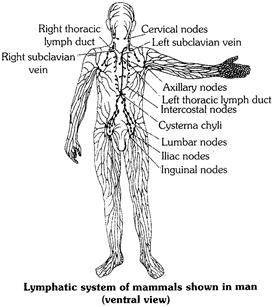
(1) Right lymphatic duct : It is the smallest lymphatic duct with the length of approximately 1.25 cm. Its one end is blind and other one opens into right subclavian vein at the junction of right internal jugular vein. It collects lymph from one-fourth of the body (right part of head, neck, thoracic cavity and right arm).
(2) Left lymphatic duct/thoracic duct : It is the longest lymphatic duct with the length of approximately 38-45 cm. It originates from cisterna chyli and empties into left subclavian vein. It collects lymph from three-fourth part of the body i.e. complete posterior part through cisterna chyli, left part of head, neck, thoracic cavity and left arms.
Cisterna chyli/Receptaculum chyli : It is a dilated sac like structure present below the diaphragm in lumbar region at the level of second lumbar vertebra. It collects lymph from posterior part of body i.e. abdomen, pelvic region and hind limbs and drains it in the left lymphatic duct.
It shows inflation and deflation due to the movement of diaphragm which is a passive movement. Hence, it is also called as passive lymphatic artery. It is also called as second heart.
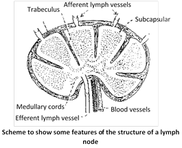
Lymph nodes or lymph glands : These are the masses of lymphatic tissue and connective tissue (reticular tissue) and are located on the capillaries either solitary or in cluster. Where they are present solitary and in few number, such tissues are called diffused lymphatic tissues and where they are in clusters, they are called tonsils.
Some of the common lymph nodes are - Axillary nodes (in armpits), genital (Inguinal) nodes (in pubic region), cervical nodes (in neck region), intercostal nodes (in chest region), lumbar nodes (in lumbar region), iliac nodes (in pelvic region) and payer’s patches (in small intestine). Besides these lymphatic nodes, a number of them are also present near major blood vessels (arteries), specially dorsal aorta.
Function of lymph nodes
(i) They produce and supply lymphocytes to the blood and as a supportive function the trabeculae carry blood vessels which supply the node.
(ii) They make screening of the lymph by means of phagocytic activity.
(iii) They serve a great defensive role against bacterial infections.
(iv) They temporarily stop the spread of cancer cells as those cells have to penetrate through the lymph vessels to the lymph nodes from where they spread in the body.
(v) They act as mechanical filters to resist the entrance of poisonous substances into circulation.
(vi) They carry out immunological responses. They help in elaboration of antibodies and in the development of immunity.
(vii) Lymph nodes produce \[\gamma -\]globulin.


You need to login to perform this action.
You will be redirected in
3 sec

