Category : 11th Class
![]()
Habitat
Funaria is known as common moss or green moss or cord moss. There are 117 species of Funaria which are worldwide or cosmopolitan in distribution. 25 species have been reported from India. Most common species are Funaria hygrometrica, F.obtusa, F. attentua and F. fasicularis.
Funaria grows well in recently burnt grounds rich in plant ashes (alkaline condition). Common places for its growth are moist rocks, moist walls and moist grounds.
External structure (Gametophytic phase) : The main plant body of Funaria is gametophyte and is of two forms.
(1) Juvenile form (creeping protonema).
(2) Adult form (leafy gametophore).
Protonema is the branched filamentous portion which is produced by germination of spores. It is ephemeral or short lived.
Leafy gametophore develop from buds produced on protonema and is made up of axis with spirally arranged leaves. It is 1 –3 cm in height green and is monopodially branched. The main branch of leafy gametophore bears male reproductive organs, i.e., antheridia and the side branch is female branch.
Leaves : These are small, sessile, ovate with acute apex and broad base. A distinct midrib is present.
On the lower portion of the leafy gametophore, there are presence of branched, multicellular rhizoids with oblique septa. Initially the rhizoids are colourless and hyaline but at maturity they become dark in colour due to dark cell walls. If rhizoids are exposed to light they become green.

Reproduction
Funaria reproduces both by vegetative and sexual methods.
(1) Vegetative reproduction : Vegetative reproduction takes place by fragmentation, primary protonema, secondary protonema, bulbils, gemma and apospory.
In apospory some cells from any part of sporophyte separate from the parent, fall on the soil and form a protonema. Buds of leafy gametophore are produced on the protonema and give rise to a gametophyte- like plant but the cells have diploid set of chromosome (2n). In this way a gametophyte like plant is produced from diploid sporophyte without reduction division.
(2) Sexual reproduction : The Funaria plants are monoecious and autoecious, i.e., male (antheridia, club shaped) and female (archegonia, flask shaped) reproductive organs are produced on the same plant but on different branches. Male organs mature first and hence Funaria plants are Protandrous.
Antheridium : Main branch is male branch which is called antheridiophore. The antheridia are borne at the tip of antheridiophore. The antheridial cluster remains surrounded by a rosette of leaves called Perigonium. In between antheridia are present club shaped green multicelled sterile hair called paraphyses. An antheridium is distinguishable into stalk and the body. It has a 1-cell thick jacket. Jacket is green in the young antheridium but turn change in mature antheridium. A number of biflagellate sperms are produced by antheridium.
Archegonium : Female receptacle appear bud like. It contains a number of archegonia intermixed with pointed 4-5 celled green paraphyses. The whole complex is surrounded by upwardly bending or convergent perichaetial leaves.
The archegonium of moss is distinguishable into a stalk, venter and a neck. The venter is double layered and contains an egg and a ventral canal cell inside. The neck is several cells high and is made up of six vertical rows of cells. It contains six or more neck canal cells. It is capped by four cover cells or lid cells. At maturity the venter canal cell and neck canal cells degenerate to form mucilaginous mass. It absorbs water and swells up and opens the lid to create a passage upto oosphere. Oosphere secretes sucrose for attracting sperms.
Fertilization : The neck canal cells and the ventral canal cell also degenerate and the cover cells split apart giving a free passage to incoming antherozoid. The fertilization is affected by water medium. Out of many antherozoids, only one of them is able to fuse with the single egg to produce zygote (2n). The zygote without any resting period enlarges and divides to form sporophyte or sporogonium. The first division in zygote is transverse. The epibasal (upper) portion forms capsule and upper half of seta while hypobasal (lower) portion forms foot and lower half of seta.
Sporogenous tissue develops from outer layer of endothecium. The venter wall forms calyptra which is present initially around the sporophyte but later on it breaks.
Sporophytic phase : Fully developed sporophyte or sporogonium is made of three regions, i.e., basal foot, seta and capsule. Sporophyte is semiparasite on moss plant taking water, minerals and some growth factors.
(1) Basal foot : Basal foot is embedded in the apex of female branch and is conical in shape. Its function is to absorb nutrients and to provide support for sporophyte.
(2) Seta : It is a long, slender, reddish brown stalk like structure which bears capsule at the top. Internally it is differentiated into a central cylinder and epidermis. The central cylinder helps in the conduction of water and the thick walled cells of the cortex provide mechanical strength to the slender seta.
(3) Capsule : Capsule is the terminal pear shaped portion. Initially it is surrounded by calyptra but later on this calyptra breaks up. Stomata are present on the lower part of capsule. The capsule consists of three portions, i.e., basal apophysis, central theca and terminal operculum.
Apophysis : Apophysis is basal sterile portion of capsule in continuation with seta. In capsule of Funaria stomata are present only in apophysis.
Theca : Theca is the fertile middle zone of capsule, situated between apophysis and operculum. The outer layer of theca is epidermis, inner to which is 2 layered hypodermis. Central sterile portion of theca is called columella, outer to which is spore sac containing spores. Columella is the supporting tissue.
Outer to spore sac there are present air spaces traversed by many portions of assimilatory cells called trabeculae.
Operculum : The upper region of capsule is slightly oblique having upper cap-like portion called operculum. Which covers the peristome. The opercular region is separated from the theca region by two rings. The lower ring is the rim or diaphragm and the upper is annulus. The peristome is distinguishable into two whorls of radially arranged peristomial teeth. In each whorl, there are sixteen teeth. Each tooth is a triangular structure. Outer ring of these teeth called exostome (hygroscopic in nature) and inner ring is called endostome (non-hygroscopic in nature).

Dehiscence of capsule : As the capsule matures the thinwalled cells including columella dry up. The thin walled cells of operculum break away. The operculum is thus separated along the annulus exposing the peristome. The hygroscopic action of the peristomial teeth also help in the removal of operculum. The capsule becomes inverted due to epinasty. It also shows hygroscopic action. The lengthening and shortening of peristomial teeth help in the dispersal of spores. The inner peristome acts as a sieve allowing only a few spores to escape at a time. Spores have a long viability, i.e., 8-15 years.
Germination of spores : The spore is the first cell of gametophytic generation. Each spore is spherical with two walls, outer one is smooth and coloured called exosporium and inner colourless smooth endosporium. Inside the spore is a single haploid nucleus, numerous chloroplasts and oil globules.
On approach of favourable conditions the spore absorbs water, outer thick exine ruptures and intine comes out in the form of a germ tube which is cut off by means of a septum (oblique). Then it divides and enlarges to form a branched alga like, filamentous, primary protonema.
There are two generations in life cycle of Funaria, i.e., gametophytic generation (n) which is independent and complex and sporophytic generation (2n) which is partially dependent upon gametophytic generation. These two generations follow each other in regular sequence. This is called heteromorphic or heterologous alternation of generations.
![]()
Habitat
It is a cosmopolitan genus. The plants occur on moist soil or rocks. Most of the species are terrestrial (on damp soils) except Riccia fluitans, which is aquatic in nature.
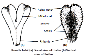
External structure (Gametophytic phase) : The main plant body of Riccia is gametophytic (n). It is small, green, flat and fleshy. The thallus is dorsiventral and dichotomously branched. The thalli are present in the form of patches called rosettes. Scales are found on the margins, while rhizoids are present in the mid-rib region of thallus. Rhizoids are unicellular and unbranched and are of two types – smooth and tuberculate. Rhizoids help in fixation. In submerged species, (e.g., R. fluitans) scales and rhizoids are not present.
Internal structure : The thallus is internally differentiated into :
An upper or dorsal photosynthetic region : It consists of vertical rows of chlorenchymatous cells. In between these vertical rows are present very narrow air canals or air chambers. The canals communicate with the outside through air pores. The uppermost cell of each row is enlarged and non-green. These non green cells of vertical rows form a discontinuous and poorly-defined upper epidermis.
A lower or ventral storage region : The lower portion consists of closely packed parenchymatous cells without intercellular spaces. The cells do not contain chloroplasts. They store water and food. The lowermost row of cells form the lower epidermis. Rhizoids and scales develop from the lower epidermis.
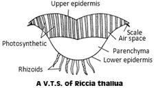
In R. fluitans and R. crystallina, the photosynthetic region is made up of chlorophyllose photosynthetic lamellae running in various directions. They enclose tiny air chambers in between them.
Reproduction
Riccia reproduces by both vegetative and sexual method.
(1) Vegetative reproduction : Riccia reproduces vegetatively by progressive death and decay, persistent apices (R. discolor), adventitious branches (R. fluitans), tubers (R. billardieri, R. discolor, R. perennis) and by rhizoid (R. glauca).
(2) Sexual reproduction : Sexual reproduction is oogamous type in Riccia. Antheridia and archegonia are the male and female sex organs respectively.
Most of the species are monoecious or homothallic, i.e., male and female sex organs are present on the same thallus. A few species are dioecious or heterothallic, i.e., antheridia and archegonia are present on different thalli. Common dioecious species of Riccia are R.himalayensis and R. frostii.
Antheridia produce biflagellated elongated curved sperms, both flagella are alike (whiplash type). Archegonia are flask shaped with neck and venter. Neck enclose 4-6 neck canal cells. Venter wall is single layered and encloses one venter canal cell and one egg cell (oosphere). It attracts sperm by secreting protein and K+ salts (chemotaxis).
Fertilization : The fertilization is affected by water medium (zooidogamous). Many antherozoids may enter into the archegonium, but only one of them is able to fuse with single egg to form zygote (2n), which is beginning of sporophytic phase.
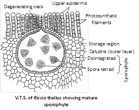
Sporophytic phase : Sporophyte or sporogonium is also embedded. It is covered by two layered calyptra. Sporogonium is undifferentiated and function like a spore sac on capsule. Nurse cell, wall of sporophyte and inner layer of calyptra degenerate to provide nourishment to growing spores. Mature spores are liberated when the surrounding cells decay or dry up.
Germination of spores : Spores are the first cells of the next gametophytic generation. Spores are dispersed by the decay of the surrounding thallus tissue. The wall of the spore is thick and sculptured, and is differentiated into three layers- the outer exosporium, the middle mesosporium and the inner endosporium which is made of pectose and callose. The surface is having clear triradiate mark. In the mass of cytoplasm, stored food is present in the form of oil – globules. After liberation, the spores germinate in about 6-10 days in presence of light, low temperature and sufficient moisture contents. After absorbing water, the spore swells up. The endosporium grows out in the form of a germ tube which, after further divisions, develops into a new thallus (gametophyte).
Thus there are 2 generations in life cycle of Riccia. The main plant body is gametophytic (n). The gametophytic phase starts with formation of spores and ends with fertilization. The second phase is sporophytic phase (2n), which starts with zygote and ends with reduction division of spore mother cell. The sporophytic phase is dependent upon gametophyte. Thus there is heteromorphic or heterologous alternation of generations in Riccia. So life cycle in Riccia is diplohaplontic.


You need to login to perform this action.
You will be redirected in
3 sec

