Category : 11th Class
(1) Axial endoskeleton : (Skull + Vertebral column + Sternum + Ribs)
(2) Appendicular endoskeleton : (Girdle + Limb bones)
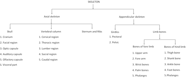
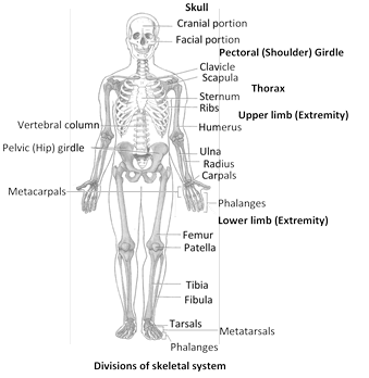
Axial skeleton (Human)
It occupies the body's main longitudinal axis. It includes four structure : skull in the head, vertebral column in the neck, trunk and tail if present, sternum and ribs in the thorax. It form the upright axis of body and includes 80 (87 in children) bones are as follows in man -
(1) Skull (General structure) : It is anterior most axial skeleton. It is divisible into two main parts –
(i) Chondrocranium (ii) Splanchnocranium
(i) Chondrocranium : Chondrocranium is formed by (a) brain box or cranium proper and (b) two sense capsules - Orbit or optic capsule (eye) and auditory or otic capsule (ear).
(a) Cranium proper : It is a strong and firm bony box with a helmet-like covering over the brain, called vault of skull, and a relatively thicker and stronger floor of base upon which the brain rests. Its cavity is called cranial cavity. Size of cranial cavity averages 1475 cubic centimetres \[(c{{m}^{3}})\] in adult men. At about the middle of the floor of cranium, there is a large opening of cranial cavity called foramen magnum. The brain is connected to spinal cord at this foramen. Cranium proper of mammal has following distinct zone -
(b) Sense capsule : Chondrocranium contains two sense capsule.
Optic capsule : One pair of optic or orbital capsule are present in frontal zone of chondrocranium. It is made up of 7 pairs of bones which are -
\[IPre\text{ }frontalIIPost\text{ }frontalIIIAnterior\text{ }orbital\]
\[IVPosterior\text{ }orbitalV\text{ }\text{ }Infra\text{ }orbitalVI\text{ }\text{ }Supra\text{ }orbital\]
\[VIILacrymal\]
In frog optic capsules are absent but in place of optic capsule eye-orbit are present in same position. In between two eye capsule, a separating bone is present in mammals only. This separating bone is called inter-orbital septum. This septum is absent in frog between two eye orbits.
Auditory or Otic capsule : Auditory capsule located between occipital and parietal zone. It has two parts – Tympanic bulla and External auditory meatus. Auditory capsule in vertebrates is formed by 5 pairs of otic bones.
(I) Preotic (II) Epiotic (III) Opisthotic (IV) Sphaenotic (V) Pterotic
Out of these 5 pairs only I pair i.e. preotic participate in formation of auditory capsule of frog i.e. amphibian. In mammals e.g., rabbit I, II & III pair fuse to form a fusion bone called periotic, which forms the auditory capsule. In reptiles and birds (aves) all 5 pairs bone combinedly constitute auditory capsule. Membranous labyrinth is enclosed in the preotic and tympanic bulla. Auditory capsule has two distinct part – Outer spongy part called patrus part and Inner bony part called mastoid part.
(ii) Splanchnocranium : It is also known as facial. It includes following parts –
Mandibular arch (I pair of visceral arch) : It is made of two arches one is upper forming upper jaw and second is lower called lower jaw. In tadpole stage upper jaw i.e. upper part of mandibular arch is formed by the fusion of three cartilage called palatine, pterygoid and quadrate. These all fused to form palato-pterygoquadrate.
Lower jaw or II part of mandibular arch is cartilagenous initially and is called Meckel's cartilage which soon changes into bony structure.
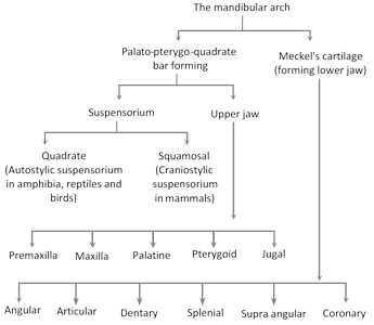
(a) Upper jaw : The upper jaw is made of 14 bones i.e. 7 pairs of bones which are - Premaxilla, Maxilla, Jugal, Squamosal, Pterygoid, Palatine, Quadrate.
Out of these 7 pairs of bones only quadrate are not visible because they constitute II ear ossicle i.e. - incus. In man the nasal cavity is separated from the buccal cavity by bone called palatine complex. Palate of birds is identical in animal kingdom, which is used for birds classification.
Process of upper jaw
Premaxilla : Nasal process on dorsal side which are covered by Nasal; Palatine process of premaxilla.
Maxilla : Nasal process of maxilla; Palatine process of maxilla; Zygomatic process of maxilla.
Squamosal : Only zygomatic process of squamosal.
Lower jaw : It is composed of 6 pairs of bone i.e. 12 bones maximum. These are articular, angular, splenial, dentary, coronoid and supra angular.
In frog out of 6 pairs only 4 pairs of bones are present. Only 3 pairs form lower jaw and one pair forms I ear ossicle i.e. collumella aures. Remaining 3 pairs i.e. Angular, splenial and dentary combine to form lower jaw of frog. In mammals only one pairs of bones are present of which only one pair i.e. dentary from lower jaw.
Upper jaw in vertebrate is completely ossified with skull but lower jaw is always free from chondrocranium and hangs downwardly. A bone hangs lower jaw from upper jaw. This bone is called suspensorium. A skull in which suspensorium is formed by quadrate is called autostylic skull e.g., frog skull. A skull in which suspensorium is formed by squamosal is called craniostylic skull e.g., rabbit skull (all mammal).
Hyoid arch (II pair of visceral arch) : It is also one pair of which is called Hyoid proper and Hyomandibular.
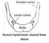
(a) Hyoid proper : It is a horse-shoe shaped bone in our neck between lower jaw and sound box or larynx. It is not articulate to any bone but is simply suspended from temporal bones by means of ligament. It consists of an elliptical main part or body and two processes on each side of body, called greater and lesser cornua. It supports our tongue and provides insertion to some tongue muscles. In colloboration with branchial arches forms hyoid apparatus in terrestrial vertebrates. It is absent in fishes because branchial arches form gill rackers which support gills.
(b) Hyomandibular : It is second part of hyoid arch which constitutes ear ossicles in vertebrate. In frog hyomandibular forms stapidial plate which is IIear ossicle which is dot or lid like bone. In rabbit hyomandibular forms stapes which is III ear ossicle. That is stirrup like bone.
Ear ossicles
|
I |
II |
III |
|
Malleus |
Incus |
Stapes |
|
Articular |
Quadrate |
Hyomandibular |
|
Hammer |
Anvil |
Stirrup |
Branchial arches (III to VII pair of Visceral arches) : These are five pairs, which constitute III to VII pair of visceral arches. These constitute gill racker in fishes but terrestrial animals then form hyoid apparatus in collaboration with hyoid proper. Five pairs of branchial arches are as follows -
(a) III pair ceratohyle.
(b) IV pair i.e. is epihyal.
(c) V pair i.e. is stylohyal.
(d) VI pair i.e. tympanohyal.
(e) VII pair i.e. thyrohyal.
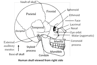
Skull of man
In man however the skull remain erect at top of vertebral column because of perfectly erect posture of body it is divisible into the large and hollow cranium and the facial region together protect and support so use organ for vision, taste, smell, hearing and equilibrium.
(1) Cranium (Brain case) : Cranium Stabilize the position of brain. In skull of man all eight bones are articulated with each other to form the cranium as follows -
Bones of cranium
|
Name |
No. |
Description |
|
Frontal |
1 |
Forms the forehead (anterior or front part of the top of cranium) and some upper parts (roofs) of eye orbits or sockets and nasal cavities. A newborn infant displays a faint suture in midline of frontal, indication that adult frontal is actually formed of two completely fused frontal. Frontal suture between two frontal disappear by age 6 years. If persists throughout life referred as metopic sutures. |
|
Parietals |
2 |
Articulated to and situated just behind frontal. Form the main parts of bulging top and sides of cranium. |
|
Occipital |
1 |
Articulated to and situated just behind parietals. Forms posterior (back) and lower (base) parts of cranium. Foramen magnum is a large perforation in this bone. On each side of the foramen, the occipital bears a prominent elevation called occipital condyle. The condyles articulate the skull with first vertebra (atlas). Thus, human skull is dicondylic. |
|
Temporals |
2 |
Form lower parts of right and left sides of cranium, as well as, the floor of cranial cavity. These house structures of internal and middle ears and form a part of external auditory meatuses. The middle ear of each side encloses the three small ear ossicles - malleus, incus and stapes. The mastoid process with mastoid air cells in adult. |
|
Sphenoid |
1 |
A typically butterfly-shaped bone that forms the middle and anterior parts of base of cranium in front of occipital in the middle and temporals on the sides. It articulates with all skull bones, keeping these firmly together. It also forms parts of lateral walls and floors of eye orbits. Sphenoid with sella turcica depression for pituitary body. |
|
Ethmoid |
1 |
A small, irregular bone in front of sphenoid and behind nasal bones. It fashions the front (anterior) extremity and closer of cranial cavity. It also contributes to the architecture of eye orbits and proximal parts of nasal chambers. |
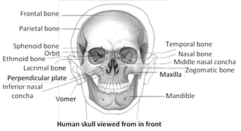
(2) The facial region : This is the front or anterior part of our skull comprised of 14 bones as follows :
Facial bones
|
Name |
No. |
Description |
|
Nasals |
2 |
Small, oblong bones in middle of upper part of face, forming proximal part of the bridge of our nose. The remaining, lower part of our nose is formed of cartilage. |
|
Inferior nasal conchae (Turbinales) |
2 |
Two highly coiled, scroll-like processes of ethmoid bone, called conchae project into each nasal cavity from lateral wall of the proximal bony part of concerned nasal chamber. One ethmoidal concha is superior (uppermost). The other one is called middle concha, because it is followed by a thin, separate scroll-like bone which is named inferior nasal concha or turbinate. |
|
Vomer |
1 |
A thin, elongated, platelike bone, forming a part of the septum which separates the two nasal cavities. |
|
Lacrimals |
2 |
Small and thin, finger-shaped bones, each located in front part of the medial (inner) side of corresponding eye orbit. these form a part of the passages of corresponding tear ducts. |
|
Zygomatics (Malars) |
2 |
Cheek-bones; form the prominences of our cheeks and parts of the floor and side walls of eye orbits. |
|
Palatines |
2 |
L-shaped bones that form the back (posterior) part of our hard palate (roof of mouth). Also contribute to the framework of nasal cavities and floor of eye orbits. |
|
Maxillae |
2 |
Large, upper jaw bones that form the major part of our face and upper jaw. Comprise entire front (anterior) part of our hard palate. Also contribute to the architecture of eye orbits and nose. Bear the teeth of upper jaw. |
|
Mandible |
1 |
Largest bone of our face, and strongest of all bones of the body. Forms entire lower jaw and bears all lower jaw teeth. Articulated with temporal bones of skull. Only skull bone that moves. |
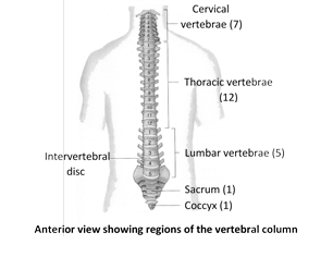
Vertebral column
It is our backbone which extends in the mid axis of the back (posterior) part of our trunk from head to the lower (inferior) extremity of trunk. Together with the sternum and rib, it forms the supporting frame work of our trunk. It support and rotate the head, suspends the viscera, protect vital organs, provides attachment to limb girdles, facilitates some movement of the trunk and houses the spinal cord. Vertebral column make two-fifth of total weight of body. The length of human vertebral column is 71 cm. (28 Inc.) in adult male and about. 61 cm (24 m.) in an average adult female.
Curvatures of vertebral column : In a foetus, there is only a single anteriorly concave curve, in adult there are 4 curves like, cervical, thoracic, lumber, and sacral. Cervical and lumber are anteriorly convex, while thoracic and sacral are anteriorly concave. At approximately 3rd month after birth, when an infant begins to hold its head erect, cervical curves develops. Later, when the child sits up, stands, and walks, the lumber curves develops. The thoracic and sacral curves retains anterior concavity of foetus thus are called ‘Primary curves’. The cervical and lumber curves are modification of the original foetal curves, are called “Secondary curves”.
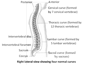
The curves of vertebral column are important because they increases its strength, help maintain balance in upright position absorb shock during walking and running and help protect the column from fracture. Certain abnormalities of curvature are:
(i) Kyphosis : Exaggeration of thoracic curve, resulting in “round-shouldered” appearance, also called hunch back.
(ii) Lordosis : An exaggeration of lumber curve, also called sway back.
(iii) Scoliosis : An abnormal lateral curvature in any region of spine.
The vestegeal notochord called nucleus pulposes is found in intervertebral disc. Inter-vertebral disc is fibro cartilagenous disc present between centrum of vertebrate.
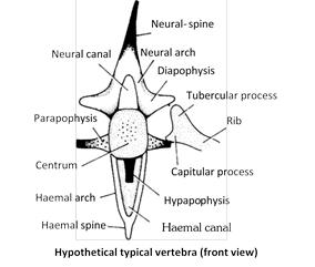
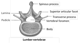
Structure of Typical vertebrae
(1) Neural arch : It arises from the dorsal side of the centrum and encloses a neural canal for the spinal cord. The arch may be produced into a dorsal process, the neural spine, which may be elongated pointed or flattened and directed upwards or backwards.
(2) Transverse processes : These are lateral extension of neural arch and centrum. There may be two types of these processes a more dorsal diapophysis arising from the base of neural arch and a lateral parapophysis arising from the side of the centrum.
(i) Diapophysis (dia- two; apo- from; physis – growth) : These paired processes are directed differently and provide attachment to the tubercular processes of ribs. They are commonly known as transverse processes and are found in amphibians and other higher vertebrates.
(ii) Parapophysis : These paired outgrowths are similar to diapophysis and are common in fishes.
(iii) Zygapophyses : These are paired and flat articular surfaces, which check the dislocation of the vertebrae. These are the only structures which enable to identify the anterior and posterior faces of vertebra.
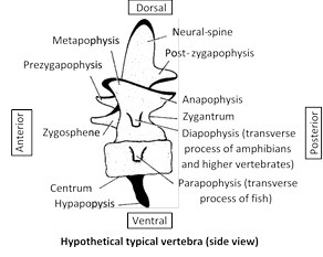
(iv) Hypapophysis : It is a mid-ventral process which arises from the centrum. It may be directed forwards or backwards as in certain reptilian, avian and mammalian vertebrae.
(v) Metapophyses : These are paired swellings or out growths having broad base and arising from just above the prezygapophyses. They are directed forwards and upwards, but their articular facets look slightly downwards. They are found in certain mammalian vertebrae.
(vi) Anapophyses : These are paired, slender and short processes which arise just below the postzygapophyses. Their articular facets look-slightly upwards and receive for the matapophyses.
(3) Heamal arch : It surrounds the haemal canal which allows the blood vessels of the tail region to pass. It may be produced into a haemal spine below e.g., in fishes. The haemal arch of the caudal vertebrae of reptiles is called chevron bone. It is usually Y-shaped.
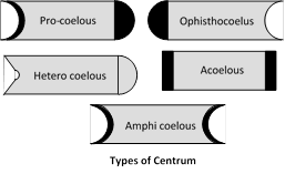
(4) Centrum : The part of vertebra attached to second by centrum. On the basis of centrum vertibrae may be of following type -
(i) Procoelus-vertebrae : Anterior end is concave and posterior end is convex. e.g., 2nd to 7th vertebra of frog. Reptile (Lizard)
(ii) Ophisthocoelus vertebrae : Anterior end is convex and posterior end is concave. e.g., Fishes, snake and crocodile only.
(iii) Heterocoelus vertebrae : Anteriorly convex from dorsal to ventral and concave from side to side. On posterior side concave from dorsal to ventral and convex from side to side (saddle shaped). e.g., Birds.
(iv) Acoelus vertebrae : Also known as Amphiplatyon. No cavity in centrum so centrum is flat. e.g., Mammals (man, Rabbit).
(v) Amphicoelus vertebrae : Cavity present on both side of centrum. e.g., VIIIth vertebra of frog. All veretebrae of scoliodon (Dog fish)
(vi) Amphidicondylar (Biconvex) : Biconvex, condyle on both side. e.g., IXth vertebra of frog.
Vertebral column of man : Made up of pieces of bones known as vertebrae. Vertebrae of man are acoelus i.e. Centrum is flat and without cavity (Amphiplatyon). Vertebral column also known as spinal column or backbone.
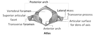
(1) Atlas vertebra
(i) First cervical vertebra.
(ii) Body is formed of vertebral arch transverse process.
(iii) It supports the globe of the head like the earth by the atlas (super man).
(iv) Centrum is absent.
(v) Neural spine absent.
(vi) Transverse process are long with transverse foramen.
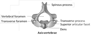
(2) Axis vertebra
(i) Second cervical vertebra.
(ii) Centrum acoelus.
(iii) Odontoid process or dens present, which is modified centrum of Atlas.
(iv) It is pivot for rotation of atlas and head around odontoid process. Transverse process small.
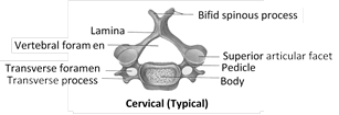
(3) Typical cervical vertebra
(i) Long neural spine.
(ii) Centrum acoelus.
(iii) Transverse process are large.
(iv) Vertebrarteal canals present.
(v) Vertebrarteal canals also known as foramina transversaria.
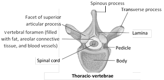
(4) Thoracic vertebra
(i) Centrum acoelus.
(ii) Neural canal is formed by union of two neural arches.
(iii) Neural spine is a flat & long directed backward.
(iv) Club shaped transverse process.
(v) Neural arch with superior articular process.
(vi) Two demifacets for articulation of head of a rib are present.
(5) Lumber vertebra
(i) Centrum acoelus.
(ii) Neural spine well developed.
(iii) Transverse process are thin and long.
(iv) Small accessory process present near the root of each transverse process.
(v) It is the largest vertebrae.
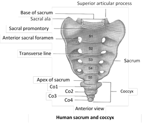
(6) Sacrum : It is a triangular bone formed by fusion of 5 sacral vertebra.
(i) Fusion normally begins between 16 to 18 years of age and is completed by 30 yrs. of age.
(ii) Serves as strong foundation for pelvic girdle.
(iii) Sacrum with 4 pairs of anterior and posterior sacral foramina.
(iv) Lateral part of sacrum articulate with ilium of hip bone.
(v) Female sacrum is shorter, wider and more curved between S2 and S3 the male sacrum is longer, narrower, and less curved.
(vi) In birds some of the vertebrae are fuse to form synsacrum. [Last thorasic + Lumber+ Sacral + One or two caudal]
(7) Coccyx
(i) It is formed by fusion of four coccygeal vertebrae.
(ii) It is last section of backbone.
(iii) It is small triangular bone.
(iv) Two coccygeal cornua project up to articulate with sacral cornua.
(v) Rudimentary transverse process.
(vi) Fusion generally occurs between 20 and 30 years of age.
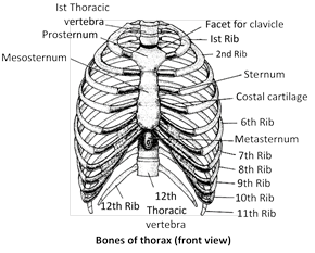
Thoracic basket
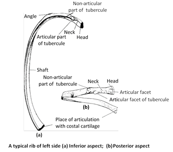
(1) Ribs
Structure : The ribs are curved bars, which movably articulate with the thoracic vertebrae at the back and while with the sternum in front all collectively forming a bony cage, the thoracic basket. These are 12 pairs of ribs. The upper seven pairs of ribs are attached in front directly to the sternum by hyaline cartilage. These are called true rib. The next three pairs of rib costal cartilage attach indirectly to sterum. They are termed false ribs. The lower two pair of ribs are free in front they are known as floating ribs. Tenth rib is also usually floating in Japanese and some other people.
A rib consists of two parts, Vertebral and Sternal. The vertebral part is long and bony. It articulate with the thoracic vertebrae by 2 facets, the capitulum and tuberculum, (Ribs of mammal and birds are bicephalous) in the first nine ribs and by a single facet, the head in the remaining vertebrae. The sternal part is short and cartilaginous. It articulate with the sternum or sternal part of its upper rib.
Human thorax is wider from side to side then from front to back. This is an adaptation for the up right posture of the body. It help to maintain equilibrium. In birds a uncinate process is present in ribs for muscles attachment.
Function : The ribs serve three important functions -
(i) They protect the heart, large blood vessels and lungs.
(ii) They bear respiratory muscle (external and internal intercostal muscle).
(iii) Lower two pair of ribs protect the kidney. (11th and 12th )
(2) Sternum
Structure
(i) It is bone of chest.
(ii) It is absent in fish, Turtle.
(iii) It is associated with pectoral girdle in amphibia.
(iv) In man it is made up of cervical manubrium (presternum), mesosternum and xiphoid process (Metasternum).
(v) In male it is nearly 17 cm long.
(vi) Manubrium is broad and thick.
(vii) Mesosternum is made up fine sternabae.
(viii) Metasternum is represent by xiphisternum which is smallest broad and thin. In mammal a cartilagenous plate attached with xiphisternum known as xiphoid cartilage (hyaline).
Function : The sternum has two function -
(i) It takes part in the formation of the protective thoracic basket.
(ii) It plays a role in the respiratory mechanism.
Appendicular skeleton
It forms the bony frameworks of limbs and their supporting girdles, and includes 126 bones as follows –
|
1. Upper extermities |
2. Lower extermities |
|
(ii) Upper limbs (arms) 60 |
(i) Pelvic girdle 2
(ii) Lower limbs 60 |
Girdles : The girdle give articulation to the limb bones. There are two types pectoral girdle (shoulder girdle) and pelvic girdle (Hip girdle). Each girdle is made up of similar right and left halves (os innominate).
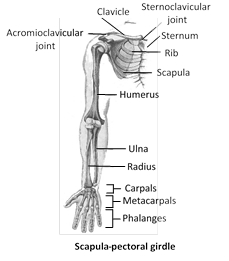
(1) Pectoral girdle
Structure : Each half (Os innominate) of the girdle mainly comprises a large, flattened and triangular cartilage bone, the scapula (shoulder blade). The broader side has a narrow strip of cartilage called suprascapula. The dorsal surface of scapula has a median longitutinal ridge called acromian spine, which successively becomes more and more prominent towards the narrower end of scapula and then, projects beyond this end as a distinct acromian process. Another prominent metacromian process projects horizontally from the base of acromian process. At its narrow end, the scapula is itself fused with an inwardly bent, knob like coracoid process. A deep, cup like concavity the glenoid cavity is located at the end of scapula close to coracoid process. The head of humerus (bone of upper arm) fits in to this cavity. Another component of each half of pectoral girdle is a long and slender, rod-like membrane bone the clavicle, articulated with the acromian process. The other end of clavicle is connected with pre sternum by means of an elastic ligament. Clavicle also called collar bone.
Function : The pectoral girdle serves two functions -
(i) It provides articulation to the arm bones.
(ii) It affords attachment to certain muscles of the arm.
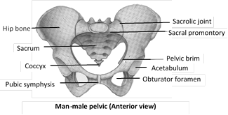
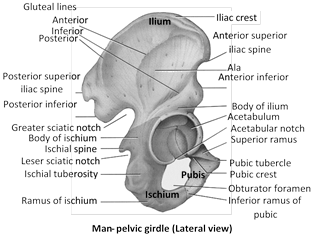
(2) Pelvic (Hip) girdle
Structure : It is located in the lower part of the trunk. It consists of 3 bones - upper ilium, lower ischium and inner pubis, fused to form a stout hip bone, the innominate. Ventral wall of pubis has a small bone called catyloid. Acetabelum is formed by ilium, ischium and pubis, but in mammals pubis is replaced by cotyloid bone. Pubic symphysis is present in mammals. Below the acetabulum, the innominate has a large oval gap, the obturator foramen (ischio-pubic foramen). The two innominate bones and sacrum together from a sort of bowel, the pelvis, that supports the lower abdominal viscera. This is also an adaptation for upright posture of the human body. The female pelvis is larger and has a broader front and larger bottom opening than the male pelvis. This is an adaptation for childbirth. In man ischial tuberocity or siting bone is present in ischium.
Functions : The pelvic girdle serves the following functions -
(i) It provides articulation to the bones of the leg.
(ii) It contributes to the formation of a bowel for the support and protection of adominal viscera.
(iii) It transfers the weight of the body to the leg.
(iv) It provides the attachment to certain leg muscles.
(v) Support vertebral column.
Limb bones : Limb are two types fore limb and hind limb.
(1) Bones of fore limbs
Structure
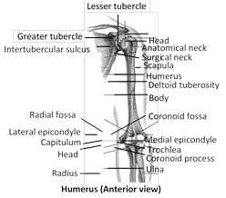
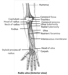
(i) Humerus or arm bone or bone of upper arm, is longest and largest bone of upper limb.
(ii) It articulates proximally with scapula and distally at the elbow with both ulna and radius.
(iii) Humerus proximal end with greater and lesser tuberosity tubercle.
(iv) Both radius and ulna with nutrient foramina.
(v) Radius present towards thumb side.
(vi) Ulna present towards little finger side.
(vii) It includes Humerus + Radius & ulna + Carpals + Meta carpals + Phalanges.
(viii) Humerus is characterised by presence of deltoid tuberocity for the attachment of muscles.
(ix) Distal end of humerus at the elbow joint is like pully and called trochlea. Its groove is called olecranon fossa whose basal part is marked by a supratrochlear foramen for the passage of brachial artery and nerve.
(x) Humerus is characterised by arterial foramen.
(xi) Head of the humerus articulate with glenoid cavity of pectoral girdle.
(xii) Radius is smaller and ulna is larger, were bones of fore arm.
(xiii) Styloid process is present in distal end of ulna and radius both.
(xiv) Olecranon process is present in ulna. Proximally, which forms prominence of elbow.
(xv) Trochlear notch is formed by ulna which is also known as sigmoid notch.
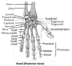
(xvi) Carpals or wrist bone are eight in number, joined to one another by ligaments. Carpals are arranged in 2 rows, with 4 bones in each row.
(xvii) Metacarpals are five in number, and phalanges are – fourteen, phalanges formula = 2, 3, 3, 3, 3.
Special features : In the human arm, (i) The joints are more movable than in the forelimbs of animals (ii) Metacarpals form a wide palm and (iii) Thumb is opposable. The differences in structure are correlated to the differences in function. Animals use their forelimbs mainly for locomotion whereas man uses the arms for work (grasping).
Function : Bones of the arms provide strength to make the arms effective in working with them.
(2) Bones of hind limbs
Structures
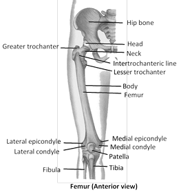
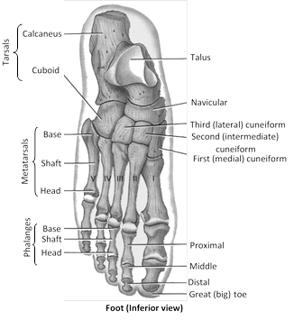
(i) It includes Femur + Tibia and Fibula + Tarsals + Metatarsals + Phalanges
(ii) Fovea capitis is depression in head of femur.
(iii) Femur is longest and strongest bone of body.
(iv) Femur is known as bone of thigh
(v) Greater trochenter, lesser trochenter 3rd trochonter are present in femur, of thigh and buttock muscles.
(vi) Patellar groove in found in distal end of femur.
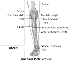
(vii) Fibula is smaller and associated with knee joint.
(viii) Tibia is larger, also called shin bone. It bears a weight of body.
(ix) Tarsal bones are seven.
(x) Metatarsals are five.
(xi) Phalanges are fourteen.
(xii) Phalanges formula\[=2,3,3,3,3\]
(xiii) Patella form knee cap.
(xiv) Patella is formed by sesamoid bone. Fabella also example of sesamoid bone.
(xv) Thumb of foot is called hallux.
(xvi) Ankle bones have 7 tarsals and arranged in two rows then Ist row have talus and calcaneus, second row with cuboid, Navicular, and I, II, III cuneiform.
(xvii) Nutrient foramen present in Tibio-fibula bone. Tibia fibula is longest bone in frog.
Special features : All the bones of the legs are more massive than the bones of the arms because the legs alone support the body on the ground and are used in locomotion. The broad feet provide an additional stable support in the upright posture.
Function : The bones strengthen the legs to bear body weight, to balance the body while standing and to aid in locomotion.
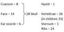
Total number of skeletal bones : 206 Bones
|
Cranium : 8 Bones Occipital : 1 Bone Parietal : 2 Bones Frontal : 1 Bones Temporal : 2 Bones Sphenoid : 1 Bone Ethmoia : 1 Bone
|
Nasals : 2 Bones Vomer : 1 Bones Turbinates : 2 Bones Lacrymal : 2 Bones Zygomatic : 2 Bones Palaline : 2 Bones Maxilla : 2 Bones Mandible : 1 Bones |
|
Coccyx : Fusion of 4 coccygeal vertebrae In new born baby : 5 sacral vertebrae In adult : Only one sacrum Ribs in man : 12 pairs True ribs : 7 pairs False ribs : 3 pairs Floatting ribs : 2 pairs |
Vertebral formula = 33 (child) C TH L S C \[\downarrow \] \[\downarrow \] \[\downarrow \] \[\downarrow \] \[\downarrow \] 7 12 5 5 4 Sacral coccygeal Vertebral column = 26 (In adult) C TH L S C \[\downarrow \] \[\downarrow \] \[\downarrow \] \[\downarrow \] \[\downarrow \] 7 12 5 1 1 Sacrum coccyx |
|
Malleus : 2 Bones Incus : 2 Bones Stapes : 2 Bones Hyoid : 1 Bone
|
Vertebral column : 26 Bones Sternum : 1 Bone Ribs : 24 Bone Pectoral girdle : 4 Bones Pelvic girdle : 2 Bones Fore limbs : 60 Bones (both) Hind limbs : 60 Bones (both) Total : 206 Bones In child : Bones 330 |


You need to login to perform this action.
You will be redirected in
3 sec

